Muscles are tissues that help the body move. This short quiz covers the key anatomical structures, clinical conditions, and drug mechanisms that most frequently appear in medical exams and clinical practice.
Start Muscle Anatomy Quiz
Results


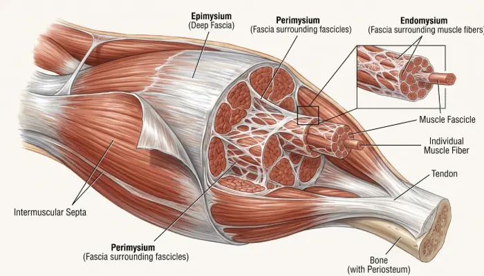
#1. Which connective tissue layer surrounds individual muscle fibers (cells) within a muscle?
The endomysium is a thin connective tissue layer wrapping each individual muscle fiber. The perimysium surrounds fascicles, and the epimysium surrounds the whole muscle.
#2. Which muscle forms the lateral border of the femoral triangle in the anterior thigh?
The femoral triangle is bounded superiorly by the inguinal ligament, medially by adductor longus, and laterally by the medial border of sartorius.
#3. Which structure connects cardiac muscle cells end-to-end and contains gap junctions for synchronized depolarization?
Intercalated discs link cardiac myocytes mechanically through desmosomes and electrically through gap junctions, allowing coordinated contraction.
#4. Which tubular invaginations of the sarcolemma carry action potentials deep into the skeletal muscle fiber?
T tubules are sarcolemmal invaginations that rapidly propagate depolarization into the fiber interior, triggering calcium release from the sarcoplasmic reticulum.
#5. Which rotator cuff muscle is traditionally described as initiating the first ~15 degrees of shoulder abduction?
The supraspinatus initiates abduction through roughly the first 15 degrees, after which the deltoid becomes the prime mover. (Newer EMG work shows supraspinatus contributes throughout the range.)
#6. Radial nerve injury in the spiral groove of the humerus causes loss of wrist and finger extension. Which classic clinical sign results?
The radial nerve supplies the wrist and finger extensors. Losing that supply produces wrist drop. The triceps is usually spared because its branches leave the nerve proximal to the spiral groove.
#7. During skeletal muscle contraction, which region of the sarcomere remains constant in width?
The A-band spans the full length of the thick myosin filaments, which do not shorten. The H-zone and I-band narrow as actin slides over myosin, and the Z-disc to Z-disc distance shortens.
#8. Myasthenia gravis is caused by autoantibodies against which component of the postsynaptic membrane?
Autoantibodies against postsynaptic nicotinic ACh receptors at the neuromuscular junction reduce signal transmission and cause fatigable muscle weakness.
#9. Succinylcholine is a depolarizing neuromuscular blocker used in rapid sequence intubation. What is its primary mechanism of action?
Succinylcholine binds nicotinic receptors at the motor endplate and, being resistant to acetylcholinesterase, causes persistent depolarization (Phase I block) and flaccid paralysis.
#10. Duchenne muscular dystrophy results from a frameshift mutation in the dystrophin gene. What is dystrophin’s role in healthy muscle?
Dystrophin connects intracellular F-actin to the dystrophin-glycoprotein complex in the sarcolemma, stabilizing the membrane during contraction.
