Cranial nerves are 12 pairs of specialized nerves that arise directly from the brain and the brainstem rather than from the spinal cord.
They carry sensory or motor nerve impulses between the brain and the structures of the head and neck, as well as other internal organs.
Quick summary table
| Nerve Number | Name | Type | Key Functions |
|---|---|---|---|
| CN I | Olfactory | Sensory | Smell |
| CN II | Optic | Sensory | Vision |
| CN III | Oculomotor | Motor | Eye movement, pupil reflex, raising eyelids |
| CN IV | Trochlear | Motor | Eye movement (downward & outward) |
| CN V | Trigeminal | Mixed | Face sensation, scalp sensation, chewing (mastication) |
| CN VI | Abducens | Motor | Eye movement (abduction/lateral movement) |
| CN VII | Facial | Mixed | Facial expressions, taste (anterior 2/3 tongue), gland secretions |
| CN VIII | Vestibulocochlear | Sensory | Hearing and balance/equilibrium |
| CN IX | Glossopharyngeal | Mixed | Throat sensation, taste (posterior 1/3 tongue), swallowing, blood pressure control |
| CN X | Vagus | Mixed | Parasympathetic control of heart & abdominal organs, throat sensation/movement |
| CN XI | Accessory | Motor | Neck movement (turning head) and shoulder elevation |
| CN XII | Hypoglossal | Motor | Tongue movements (speech, chewing, swallowing) |
They are responsible for functions such as smell, vision, hearing, taste, facial movements, eye movements, swallowing, and control of certain glands and organs.
Olfactory nerves: (sensory)
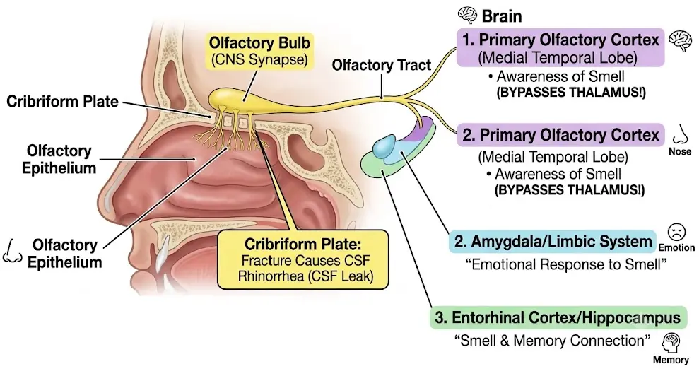
- This is the first cranial nerve, a sensory nerve responsible for the sense of smell.
- The olfactory nerves carry the sense of smell from the olfactory receptors lying in the roof of the nasal cavity to the brain.
- Their nerve fibers pass through the plate of the ethmoid bone and reach the olfactory bulb.
- The nerves then proceed backward along the olfactory tract to the olfactory bulb, the brain region responsible for the perception of smell.
Optic nerve: (sensory)
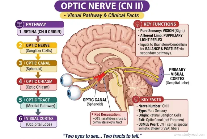
- The optic nerve is the second cranial nerve and a sensory nerve responsible for vision (sight).
- The nerve fibers originate from the retina of each eye and then combine to form the optic nerves.
- The optic nerves pass through the optic canal of the sphenoid bone into the cranial cavity.
- The visual pathways then project via the optic tracts to the thalamus and then to the primary visual cortex in the occipital lobe.
Oculomotor nerves: (motor)
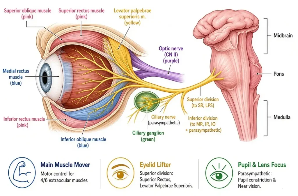
- This is the third cranial nerve.
- It is a motor nerve and controls most of the movements of the eyeball and certain muscles involved in vision, like the
- Four of six extrinsic muscles, which move the eyeball, i.e.,
- the superior
- medial
- inferior recti and
- inferior oblique muscle.
- The intrinsic muscles:
- The ciliary muscles, which alter the shape of the lens, change its refractive power.
- Circular muscles of the iris, which constrict the pupil
The levator palpebrae muscles, which raise the upper eyelids.
Trochlear nerves: (motor)
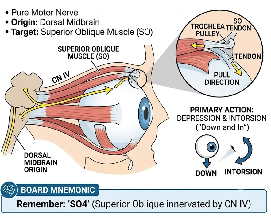
- It is the fourth and the smallest cranial nerve.
- It is a motor nerve and innervates the superior oblique muscle of the eye.
- It originates in the midbrain, at the level of the inferior colliculus.
- CN IV is the only cranial nerve that emerges from the dorsal (posterior) aspect of the brainstem and the only CN where all motor fibers intersect (cross) before exiting
- It helps the eye move downward and outward, especially when the eye is turned inward.
Trigeminal nerves: (mixed)
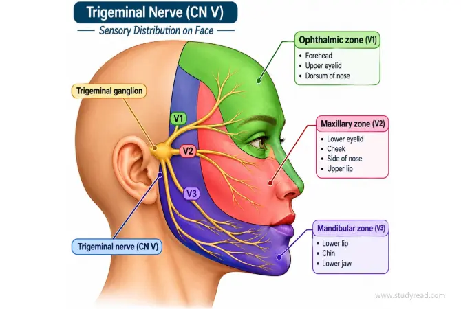
- It is the fifth cranial nerve and the largest of all the cranial nerves.
- The trigeminal nerve is a mixed nerve, with both sensory and motor fibers.
- The sensory fibers innervate the face, scalp, cornea, nasal cavity, mouth, and teeth, transmitting impulses like temperature, pain, and touch.
- The motor fibers innervate the muscles of chewing (mastication).
The trigeminal nerve has three main branches.
- The ophthalmic nerves are sensory only and supply the lacrimal glands, conjunctiva of the eyes, forehead, eyelids, and mucous membrane.
- The maxillary nerves are sensory only and supply the cheeks, upper gums, upper teeth, and lower eyelids.
- The mandibular nerves contain both sensory and motor fibers. These are the largest of the three divisions, and they supply the teeth and gums of the lower jaw, pinna of the ear, lower lip, and tongue. The motor fibers innervate the muscles involved in chewing.
Abducens nerves: (motor)
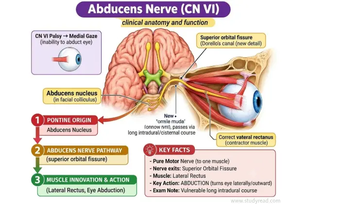
- The sixth cranial nerve, which mainly supplies the lateral rectus muscles of the eyeballs.
- This motor nerve emerges from the pons and medulla, travels forward through the skull, and enters the orbit through the superior orbital fissure.
- The innervated lateral rectus muscle moves the eyeball laterally (abduction).
Facial nerves: (mixed)
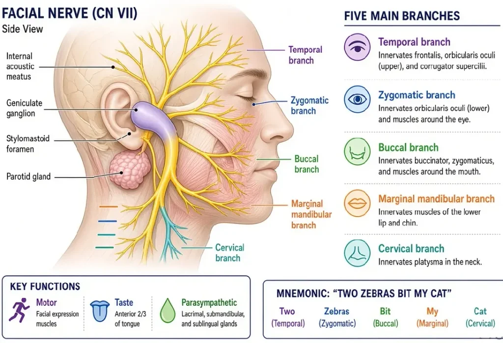
- The seventh cranial nerve is termed the facial nerve.
- It is a mixed nerve and has both motor and sensory fibers.
- This nerve emerges from the pons structure of the brain.
- The main motor function is to supply the muscles of facial expressions.
- The sensory function is to stimulate the lacrimal, submandibular, and sublingual glands.
- Taste sense from the anterior two-thirds of the tongue.
- The branches, such as Temporal, Zygomatic, Buccal, Marginal mandibular, and Cervical, mainly supply the muscles of facial expression.
| Branch | Target Muscle | Function (Tested on Exam) |
|---|---|---|
| Temporal (T) | Frontalis, Orbicularis oculi | Raising eyebrows, wrinkling forehead |
| Zygomatic (Z) | Orbicularis oculi | Tightly closing eyes |
| Buccal (B) | Buccinator, Orbicularis oris | Smiling, puffing out cheeks, blowing |
| Marginal Mandibular (M) | Depressor anguli oris | Frowning, pulling the corners of the mouth down |
| Cervical (C) | Platysma | Tensing neck skin |
Vestibulocochlear nerves: (sensory)
- Also called the auditory nerve, it is the eighth cranial nerve.
- It is a special sensory nerve concerned mainly with hearing and the maintenance of balance and equilibrium.
- The vestibular nerves arise from the inner ear and convey the impulses to the cerebellum.
- The vestibulocochlear nerve has 2 divisions:
The cochlear division is concerned with hearing.
The vestibular division is concerned with balance and equilibrium.
Glossopharyngeal nerve: (mixed)
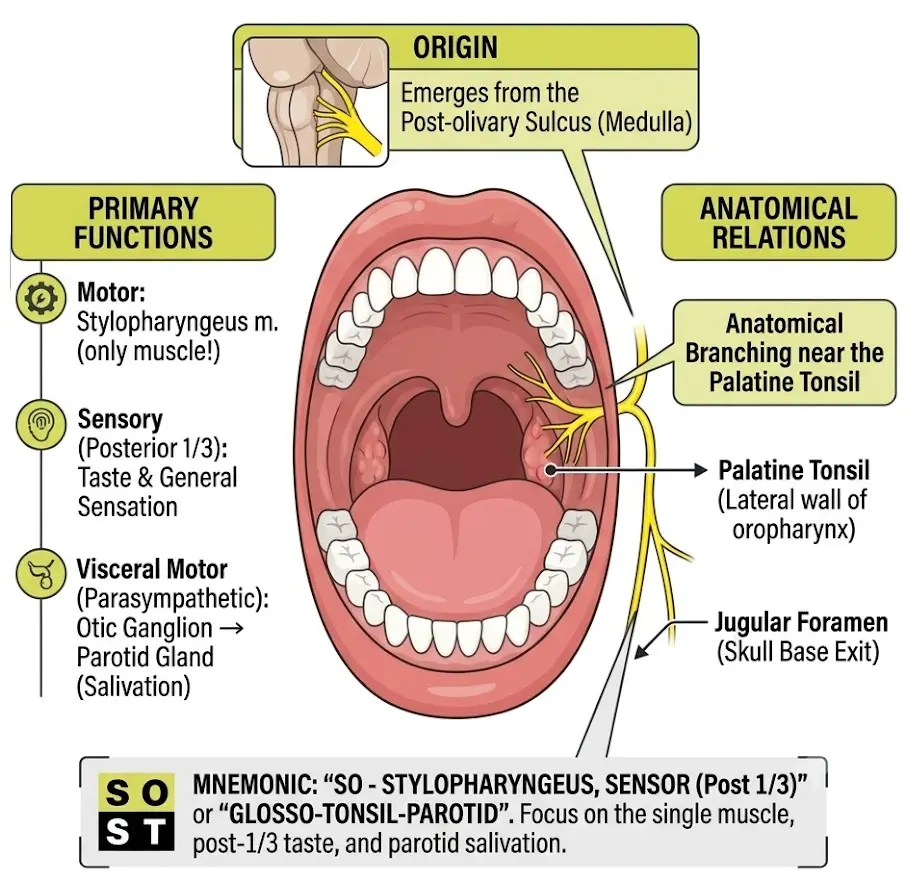
- The glossopharyngeal is the ninth cranial nerve.
- It is a mixed nerve, containing sensory and motor nerve fibers.
- The motor fibers arise from the medulla oblongata.
- The sensory fibers convey impulses to the cerebral cortex, providing sensation from the pharynx, tonsils, middle ear, and the tongue’s taste buds.
- The motor function is to elevate the pharynx during swallowing.
- Some fibers conduct impulses from the carotid sinus, which plays an important role in the control of blood pressure.
Vagus nerve: (mixed)
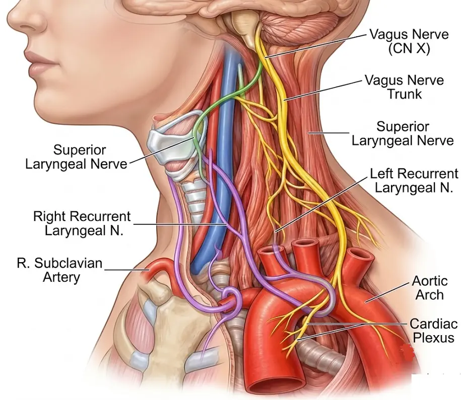
- The vagus is the tenth cranial nerve and is the longest cranial nerve.
- It is a mixed nerve containing sensory and motor nerve fibers. They pass downwards through the neck into the thorax and the abdomen.
- The motor fibers arise from the medulla oblongata and innervate the smooth muscle of the pharynx, larynx, trachea, bronchi, heart, etc.
- The sensory fibers convey impulses from the organs of the thorax and abdomen to the brain.
Accessory nerves: (motor)
- It is the eleventh cranial nerve and motor in function.
- The nerve arises from the spinal cord and the medulla oblongata.
- It innervates the muscles involved in the movement of the head, neck, and shoulder.
- The fibers supply the sternocleidomastoid, which helps turn the head to the opposite side, and the trapezius muscles, which help elevate the shoulder and assist in the movement of the scapula.
Hypoglossal nerves: (motor)
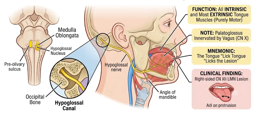
- The twelfth and last of the cranial nerves.
- It is a motor nerve that mainly supplies the muscles of the tongue.
- These nerves are essential for tongue movements involved in speech, chewing, and swallowing.
- These hypoglossal nerves arise from the nuclei in the medulla oblongata.
- The hypoglossal nerve supplies almost all the muscles of the tongue, such as the intrinsic muscles of the tongue, genioglossus, hyoglossus, and styloglossus.
References:
- Waugh, A., & Grant, A. (2022). Ross & Wilson Anatomy and Physiology in Health and Illness (14th ed.). Elsevier Health Sciences.
- Le, T., & Bhushan, V. (2023). First Aid for the USMLE Step 1 2023. McGraw-Hill Education. (Access via McGraw-Hill Medical)
- Standring, S. (2020). Gray’s Anatomy: The Anatomical Basis of Clinical Practice (42nd ed.). Elsevier.
- Gilroy, A. M., MacPherson, B. R., & Wikenheiser, J. C. (2021). Anatomy: An Essential Textbook (3rd ed.). Thieme Medical Publishers.
- Netter, F. H. (2018). Atlas of Human Anatomy (7th ed.). Elsevier.
