The heart is a muscular organ situated in the middle of the chest, slightly to the left.
It is a conical-shaped hollow organ enclosed within a pericardium, the peritoneal counterpart of the heart.
It is placed obliquely, behind the body of the sternum and costal cartilages, one-third of it to the right, and the remaining two-thirds to the left of the median plane.
It measures 9cms and weighs 300g in males and 250g in females.
Courtesy: Springer.
Anatomical Structure of the Heart
The heart looks simple, but it is an organ with a complex structure and functionality.
For convenience, we will study the structure of the heart as
External and
Internal anatomy of the heart
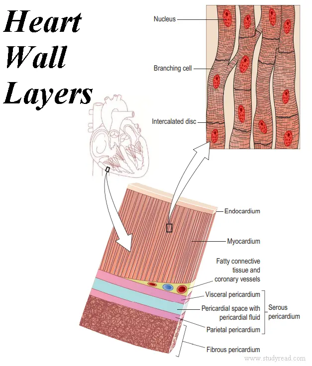
The heart wall is made up of three layers of tissue, as
It acts as a cover for the remaining structure of the heart.
Besides, it also helps fix the heart in its position in the thoracic cage.
It has two sacs as
a) Outer Fibrous sac
b) Inner Serous layer (a double membrane)
✔ The outer fibrous sac is made of fibrous tissue and is non-elastic.
It attaches the heart to the diaphragm and the rest of the surroundings and prevents its distension.
✔ The Inner layer is a continuous double layer of serous membrane, like
🌟 Outer Parietal pericardium
🌟 Inner Visceral pericardium or epicardium.
The parietal pericardium lines the outer fibrous sac.
The inner Visceral pericardium is attached to the myocardium (heart middle layer).
The cells of the serous membrane secrete the serous fluid between the two layers of the serous membrane.
This fluid in the space between the layers helps in smooth movements during the heartbeat.
Myocardium
👉This is the middle layer of the heart wall.
It consists of cardiac muscle, which is striated muscle but involuntary in nature.
The muscle fibers are branched, due to which the layer appears like a sheet of muscle.
In between these muscle fibers, there is a connective tissue that helps in the propagation of heart impulses.
Endocardium
👉This is the inner layer of the heart that forms the lining of the heart chambers.
It is made of flattened epithelial cells, which help in the smooth movement of the blood inside the heart chambers.
Internal Anatomy of the Heart
The heart is divided into the right and left sides by a septum, which is made of myocardium and covered with endocardium.
Each side is divided into the upper atrium and lower ventricle by the atrioventricular valves.
Thus, there are four chambers in a human heart. Each right and left halves contain one atrium and one ventricle.
The atria are the chambers for receiving blood, and the ventricles pump out the blood to the respective sectors.
Atria lies above and behind the ventricles.
On the surface of the heart, the ventricles are separated from the atria by an atrioventricular groove..
Further, the ventricles are separated from each other by interventricular grooves.
Chambers of Heart
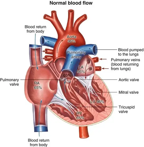
The human heart is a four-chambered organ where there is complete separation of oxygenated and deoxygenated blood.
It has two atria and two ventricles.
Right Atrium
The right atrium is the right upper chamber of the heart.
It receives venous blood from the systemic circulation, i.e. whole body, and pumps it into the right ventricle via the Tricuspid opening.
Along the right border of the atrium, there is a shallow groove that contains the Sinoatrial Node or SA Node, the pacemaker of the heart.
The right atrium is a more or less vertically elongated structure receiving the Superior Venacava from the upper end and the Inferior Venacava from the lower end.
Right Ventricle
The right Ventricle is a triangular chamber that receives blood from the right atrium.
It pumps blood into the lungs through the Pulmonary artery.
The Interior of it has two orifices –
Right atrioventricular (guarded by the Tricuspid valve) and
Pulmonary orifice (guarded by the pulmonary valve).
The septomarginal trabecula or moderator band is a muscular ridge extending from the ventricular septum to the base of the anterior papillary muscle. It contains the right branch of the Atrio Ventricular Node or AV NODE.
The Wall of the right ventricle is thinner than that of the left ventricle. The ratio of thickness is 1:3.
Left Atrium
This is a chamber located posterior to the right atrium.
It receives oxygenated blood via four branches of pulmonary veins and pumps the same into the left ventricle via the Left Atrioventricular aperture and the Mitral valve.
Two Pulmonary veins open into the left atrium on each side of the posterior wall.
In the embryonic phase, the right atrium and left atrium remain attached through an opening in the common atrial septum called the FORAMEN OVALE. Later, after birth, it closes.
Left Ventricle
The left Ventricle receives oxygenated blood from the left atrium via the mitral orifice.
It then pumps the blood into the Aorta via the Aortic valves.
It shows two orifices –the left atrioventricular guarded by the Mitral valve and the Aortic Orifice guarded by the Aortic Valve.
It is the thickest chamber of the heart, about three times thicker than the right ventricle.
Valves of the Heart
The heart is supplied with a series of valves to prevent the backflow of blood.
Classification:
The valves present in the heart are of two categories:
✔Auriculo-ventricular valves:
They are two in number, namely
Tricuspid valves: Present between the right atrium and the right ventricle (have three cusps).
Bicuspid valves: Present between the left atrium and left ventricle, it is called Bicuspid because of its two cusps. It is also known as the Mitral Valve.
✔ Semilunar Valves:
These are present between the blood vessels and the Ventricles.
Aortic valve: Present between the left ventricle and the aorta.
Pulmonary Valve: Present between the right ventricle and the Pulmonary Artery.
Circulation of blood through the body
The human body consists of two types of circulation.
Systemic circulation
Pulmonary circulation
Systemic Circulation
This circulation starts from the left ventricle, exists through the Aorta, and supplies the whole body.
The body tissues get oxygenated blood through this circulation.
This ends in the right atrium as the blood returns from the upper part of the body via the Superior Venacava, and the lower part of the body via the Inferior Vena Cava.
Pulmonary Circulation
Pulmonary circulation
This circulation starts from the right ventricle, exits the heart via the Pulmonary Artery.
From these pulmonary arteries, it reaches the two lungs, where it gets oxygenated and returns to the left atrium via the Pulmonary Veins.
The Pulmonary Artery and the Pulmonary vein are exceptional blood vessels, where an artery carries deoxygenated blood and a vein carries oxygenated blood.
Dr. Ranga Reddy N, Ph.D.
Professor of Pharmacology | IIT (BHU) Alumnus
Dr. Ranga Reddy N is a Professor and researcher with over 15 years of experience specializing in Clinical Pharmacology and Pharmaceutical Analysis. His work focuses on the intersection of drug mechanisms and clinical research. Through StudyRead, he provides evidence-based pharmacological insights for the global healthcare and scientific community.