The skin is the largest organ of the body, forming the outer protective covering.
It protects the body from injury, microbes, and dehydration, and also helps in sensation, temperature regulation, and vitamin D synthesis.
Primary Layers of Human Skin
- Epidermis
- Dermis
- Hypodermis (subcutaneous tissue)
| Layer | Primary Tissue | Main Function | Key Structures |
|---|---|---|---|
| Epidermis | Keratinized stratified squamous epithelium | Provides a waterproof barrier, protection against mechanical/chemical damage, and prevents water loss | Keratinocytes, Melanocytes, Langerhans cells, Merkel cells |
| Dermis | Dense irregular connective tissue | Gives strength, elasticity, and nourishment to the skin | Blood vessels, nerves, hair follicles, sweat glands, sebaceous glands, lymphatic vessels |
| Hypodermis (Subcutaneous) | Adipose (fat) and loose connective tissue | Thermal insulation, shock absorption/cushioning, energy storage | Adipocytes, larger blood vessels, and nerves |
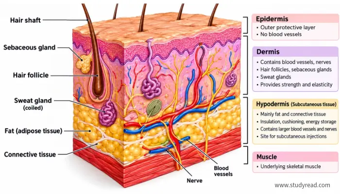
Epidermis:
- Epidermis is the outermost layer of the skin.
- It is made mainly of keratinized stratified squamous epithelium.
- It protects against mechanical and chemical damage.
Dermis:
- Dermis lies beneath the epidermis.
- It contains blood vessels, nerves, hair follicles, sweat glands, and sebaceous glands.
- Provides strength and nourishment to the skin.
Hypodermis (subcutaneous tissue):
- The hypodermis lies below the dermis and is not part of the skin.
- It mainly consists of fat and connective tissue.
- Provides insulation, cushioning, and energy storage.
- It is the tissue targeted by the subcutaneous injections.
Layers of Epidermis:
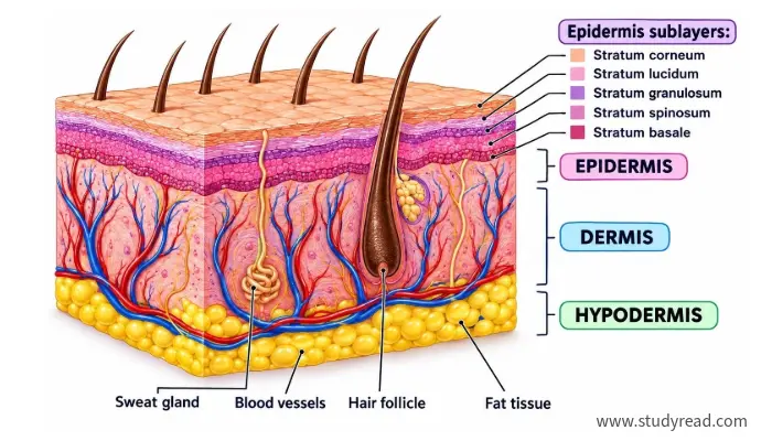
The epidermis is divided into 5 layers, arranged from superficial to deep. They are:
- Stratum corneum
- Stratum lucidum
- Stratum granulosum
- Stratum spinosum
- Stratum basale
| Sublayer | Structural Characters | Cellular Features and Functions |
|---|---|---|
| Stratum Corneum | 15–30 layers of dead, flattened, fully keratinized cells | Outermost protective barrier; continuously sheds dead corneocytes |
| Stratum Lucidum | Thin, clear band of dead keratinocytes | Present only in thick skin (palms of hands and soles of feet); provides extra protection against friction |
| Stratum Granulosum | 3–5 layers of flattened keratinocytes | Accumulates keratohyalin granules; secretes glycolipids to form a water-resistant seal |
| Stratum Spinosum | Several layers of polygonal keratinocytes | Connected by strong desmosomes (“spiny” appearance); contains Langerhans cells for immune defense |
| Stratum Basale | Single layer of cuboidal/columnar stem cells | Deepest layer; active cell division (mitosis); contains Melanocytes (pigment) and Merkel cells (touch receptors) |
Stratum corneum
- It is the outermost layer of the epidermis.
- Consists mainly of dead, flattened, keratin-filled cells called corneocytes.
- It provides the skin’s main protective barrier.
- Prevents excessive water loss and entry of microorganisms.
- Cells are continuously shed from the surface.
Stratum lucidum
- A thin, clear layer present only in thick skin, such as the palms and soles.
- It is located between the stratum corneum and the stratum granulosum.
- It contains the flattened, dead keratinocytes.
Stratum granulosum
- Contains flattened keratinocytes with keratohyalin granules.
- It is located between the stratum lucidum and the stratum spinosum.
- Keratin production increases in this layer.
- Helps form the skin’s water-resistant barrier.
Stratum spinosum
- Located below the stratum granulosum and above the stratum basale.
- Contains several layers of keratinocytes connected by strong desmosomes, giving the cells a “spiny” appearance.
- It provides the strength and flexibility to the epidermis.
- Contains Langerhans cells, which participate in immune defense.
Stratum basale
- It is the deepest layer of the epidermis.
- It consists of a single layer of actively dividing cells.
- Produces new keratinocytes that gradually move toward the surface.
- Contains melanocytes, which produce melanin and help protect the skin from UV radiation.
- It also contains Merkel cells, involved in touch sensation.
Subcutaneous vs intramuscular injection sites
Skin is the primary site for parenteral drug administration.
One can administer a drug to bypass the gastrointestinal tract by injecting the drug directly into
- Intradermal
- Subcutaneous fat
- Intramuscular (skeletal muscle below skin)
Doing so will allow the drug to enter the systemic circulation and thereby avoid first-pass metabolism in the liver.
| Parameter | Intradermal (ID) | Subcutaneous (SC) | Intramuscular (IM) |
|---|---|---|---|
| Target Tissue Layer | Dermis (below epidermis) | Hypodermis / Adipose tissue | Skeletal Muscle |
| Injection Angle | 10° – 15° | 45°-90° | 90° |
| Absorption Rate | Slowest | Slow & Sustained | Rapid (High vascularity) |
| Maximum Volume | ≈ 0.1mL | 1.0 – 1.5mL | 2.0 – 5.0mL (site-dependent) |
| Needle Specs | 26 – 27 G (3/8 – 5/8inch) | 25 – 31 G (3/8 – 5/8 inch) | 20 – 23 G (1-1.5 inches) |
| Key Clinical Uses | Mantoux TB test, allergy testing | Insulin, Heparin/LMWH, MMR vaccine | COVID-19/Flu vaccines, antibiotics, hormones |
| Primary Anatomical Sites | Inner forearm, upper back | Abdomen, outer upper arm, thigh | Deltoid, Ventrogluteal, Vastus Lateralis |
Subcutaneous injection
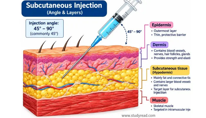
- A subcutaneous injection delivers the medication into the fatty layer of tissue located directly beneath the skin and above the muscle
Common sites of subcutaneous injection:
- Abdomen: Commonly used for injecting insulin and some other medicines.
- Outer aspect of upper arm: Fatty area over the triceps region.
- Anterior/lateral thigh: Fatty tissue of the thigh.
- Hip area: May be used for selected medications.
Characteristics:
- Uses a relatively short, fine needle.
- Absorption is generally slower than IM (intramuscular) injection.
- Suitable for medicines that need gradual absorption, such as insulin and some anticoagulants.
Intramuscular injection
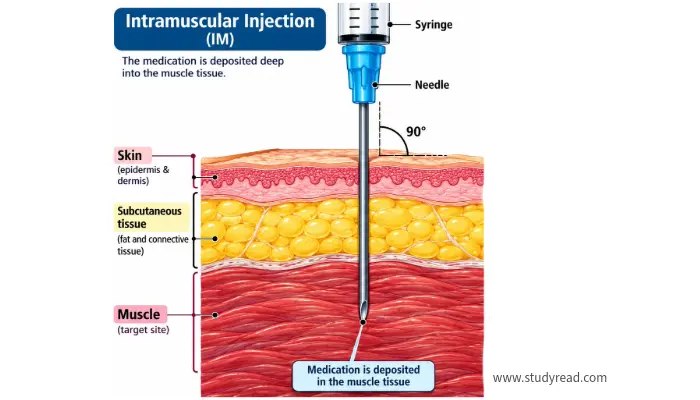
- An intramuscular injection delivers medicine directly into skeletal muscle.
Common sites of intramuscular injection:
Deltoid muscle:
- It is located in the upper arm.
- Common uses for vaccines and small-volume injections.
Vastus lateralis:
- Located on the outer side of the thigh.
- Frequently used in infants and children and can be used in adults.
Ventrogluteal site:
- Located around the hip/gluteal region.
- Considered a preferred site for many larger-volume IM injections because of its relatively safe anatomy.
Dorsogluteal site:
- Traditionally used in the buttocks, but it is generally avoided where safer alternatives are available because of the risk of injury to the major blood vessels.
Characteristics:
- Uses a longer needle than a typical SC (subcutaneous) injection.
- Absorption is usually faster with intramuscular administration than with subcutaneous administration because muscle has a good blood supply.
- The appropriate needle length depends on the patient’s age, body size, injection site, and tissue thickness.
Intradermal injection
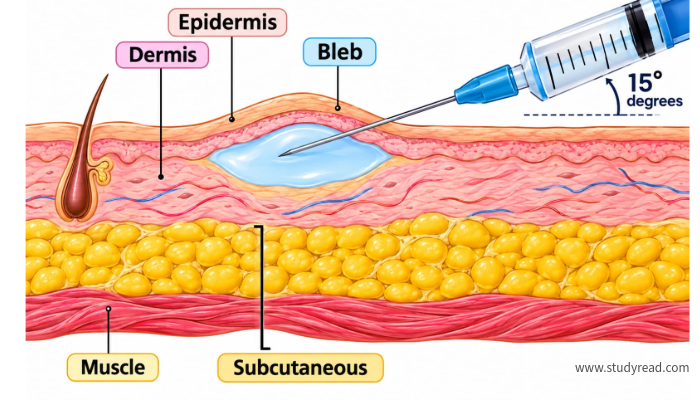
Intradermal injection is a method of administering a small volume of a drug, vaccine, or test substance into the dermis of the skin, just below the epidermis.
Common sites used for intradermal injections include:
- Inner surface of the forearm: most commonly used for skin tests.
- Upper back: used for some skin allergy tests.
- Upper chest: may be used for selected skin tests.
- Upper arm: Used for certain vaccines such as BCG, depending on the vaccination protocol.
Characteristics:
- Injection is given into the dermis of the skin.
- Uses a very small volume of solution.
- The needle is inserted at a low angle (5 – 15 degrees)
- A small raised wheal usually appears after injection.
- Absorption is slow compared with intramuscular injection.
- Mainly used for certain skin tests and certain vaccines.
- The inner forearm is a common site for skin testing.
