The brain is the principal organ that controls the human body.
It is part of the central nervous system, along with the spinal cord, and is located inside the skull. It consists of part like
| Brain Region | Major Subdivisions | Primary Clinical & Physiological Functions |
| Forebrain | • Cerebrum • Thalamus & Hypothalamus • Limbic System & Pituitary | Voluntary movement, sensory perception, language (Broca/Wernicke), memory, emotion, and endocrine regulation. |
| Midbrain | • Tectum (Superior/Inferior Colliculi) • Tegmentum (Substantia Nigra, Red Nucleus) • Cerebral Peduncles | Visual and auditory reflexes, dopamine production, motor path relays, and CSF passage via the cerebral aqueduct. |
| Hindbrain & Brainstem | • Pons • Cerebellum • Medulla Oblongata | Autonomic survival functions (breathing, heart rate, blood pressure), postural balance, and fine motor coordination. |
The main parts of the brain are
- Forebrain: Cerebrum, Thalamus, Hypothalamus, Limbic System.
- Midbrain: Tectum, Tegmentum.
- Hindbrain / Brainstem: Pons, Medulla Oblongata, Cerebellum.
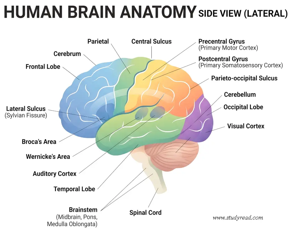
The brain controls our thoughts, speech, and memory; the locomotor movements of the hands and legs; and also the function of many internal organs.
The brain receives the information from our five sense organs:
- Eyes (sight),
- Ears (hearing),
- Nose (smell),
- Skin (touch), and
- Tongue (taste).
Using the stimulus information sent by these five sense organs, the brain makes sense of its surroundings. It is responsible for intelligence, creativity, and emotional responses.
The brain is located inside the bony cranium (skull) and is covered by meninges, which act as a cushion for protection. There are three layers of the meninges from outside to inside as
- Dura mater,
- Arachnoid mater, and
- Pia mater.
Parts of the Brain in Detail
Forebrain:
Cerebrum
- It is the largest part of the brain and consists of two cerebral hemispheres.
- The cerebrum is divided into several distinct parts separated by fissures. Each cerebral hemisphere has four lobes:
- The frontal,
- Temporal,
- Parietal, and
- Occipital lobes.
Lobes of the brain
- The lobes of the brain do not work alone. There is a complex relationship between the brain’s lobes and between the right and left hemispheres.
Frontal lobe
- Personality, behavior, judgment, planning, problem-solving, comparison, impulse control, memory formation, and empathy are controlled by this lobe.
- Further, speech, speaking, and writing are controlled by a region called Broca’s area in the frontal lobe. This helps with speaking and understanding language.
The primary motor cortex is present in the frontal lobe. It coordinates voluntary movements like walking and running by generating neural impulses.
The olfactory bulb is located on the ventral surface of the frontal lobe. It receives nerve impulses about various odors detected by the cells in the nasal cavity. Then it processes information about these odors.
Intelligence, concentration, self-awareness, motivation, and seeking behavior are all managed by this lobe. Most of the brain’s dopamine-sensitive neurons are in the frontal lobe. Dopamine is a neurotransmitter chemical that helps support feelings of motivation.
The central sulcus is a prominent fissure separating the parietal lobe and the frontal lobe. It further divides the primary motor cortex from the primary somatosensory cortex.
Parietal lobe
- The parietal lobes reside behind the frontal lobes and above the temporal lobes.
- These lobes help us understand spatial orientation and navigation. Being able to identify the position and movement of the body and its parts is an essential function of the parietal lobes.
- The somatosensory cortex is another vital region located within the parietal lobes, and it helps process touch sensations and sense pain or temperature differences.
- Parietal lobes receive impulses from the thalamus for vision, hearing, and other sensory information and help process and identify them.
Occipital lobe
- This part is the rearmost lobe of the brain. It rests upon the tentorium cerebelli, a thick membrane of tissue that separates the cerebrum from the cerebellum.
- Interprets vision (color, light, movement, depth) and transmits visual information for storage in memory.
Temporal lobe
- The temporal lobe is located just behind our ears, below the cortex, and extends to both sides of the brain.
- The main function of the temporal lobes is to process audible sounds. The primary auditory cortex is located in this lobe. It helps in the formation of sensory memories.
- The task of understanding language is performed in Wernicke’s area of this lobe.
Cortex
- This is the outer surface of the cerebrum called the cortex.
- It has a folded appearance due to the presence of neurons.
- The foldings present in the cortex increase the brain’s surface area, allowing more neurons to fit.
- A fold is called a gyrus, and the valley between them is a sulcus.
- The neurons are gray in color due to their nerve cell bodies (gray matter). Beneath the cortex are long nerve fibers (axons) that connect brain areas to each other and appear as white matter.
Deep structures
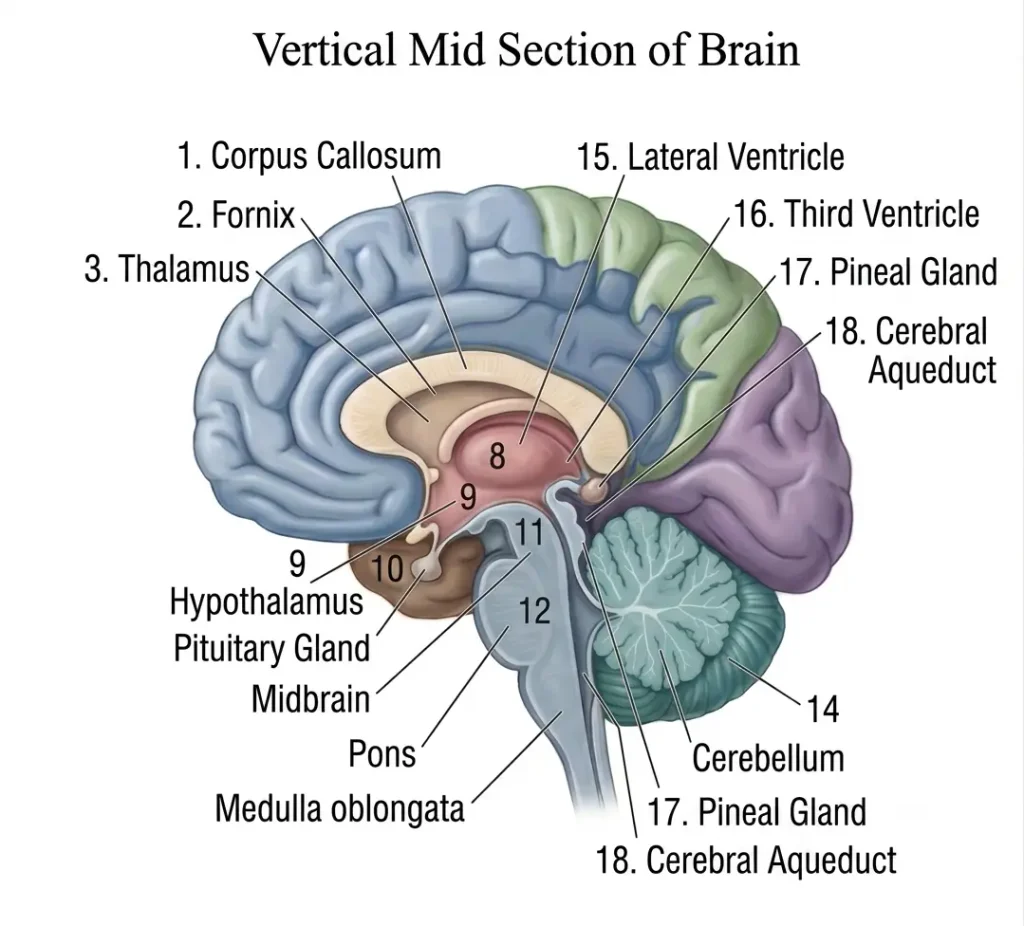
- This includes the brain’s inner structures. This consists of
Thalamus
- It serves as a relay station for almost all information that comes and goes to the cortex.
- It helps in pain sensation, attention, and memory.
- It acts as a transmission center for all the sensory information.
Hypothalamus
- It is the master control of the autonomic nervous system.
- It is positioned on the floor of the third ventricle. It regulates the secretion of hormones.
- It plays a role in hunger, thirst, sleep cycles, and sexual response.
- It also regulates body temperature, blood pressure, and emotions.
Pituitary gland
- It is located in the sella turcica, a small pocket of bone at the base of the skull.
- The pituitary gland is fixed to the bottom of the hypothalamus of the brain by the pituitary stalk.
- It is known as the master gland because it controls other endocrine glands by secreting hormones that regulate metabolism, stress, sexual cycles, and more.
Limbic system
- It is the center of our emotional responses and memory.
- The cingulate gyri, hypothalamus, amygdala (for emotional responses), and hippocampus (for memory) are all part of this system.
Pineal gland
- It is located near the thalamus, behind the third ventricle.
- It secretes melatonin and helps regulate the body’s internal clock and circadian rhythms. It also plays some role in sexual development.
Take the Brain Anatomy Quiz to test your Knowledge.
Midbrain:
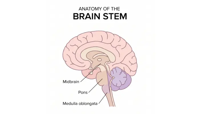
Also called the Mesencephalon, the uppermost part of the brainstem relays signals between the forebrain and the lower brainstem/spinal cord. It is structurally divided into three main regions:
- Tectum
- Tegmentum
- Cerebral Peduncles
Tectum (Dorsal Region / Roof)
- Contains the corpora quadrigemina, four rounded swellings responsible for visual and auditory reflexes.
- Superior Colliculi: Reflex centers for visual tracking and eye-head movement coordination.
- Inferior Colliculi: Reflex centers for processing auditory signals (e.g., turning toward a sudden sound).
Tegmentum (Ventral Region / Core)
- Forms the main body of the midbrain between the tectum and cerebral peduncles.
- Substantia Nigra: High density of dopaminergic neurons involved in smooth motor control (degeneration here is the primary cause of Parkinson’s disease).
- Red Nucleus: Coordinates motor output and limb movement.
- Cerebral Aqueduct: The canal passing through the midbrain that channels cerebrospinal fluid (CSF) from the 3rd to the 4th ventricle.
Cerebral Peduncles (Anterior Base)
- Nerve fiber bundles (corticospinal and corticobulbar tracts) carry motor signals directly from the cerebral cortex to the brainstem and spinal cord.
Hind Brain
Pons
- It is a part of the brainstem that connects the cerebral cortex to the medulla.
- The pons helps transmit messages within the nervous system between various parts of the brain and the spinal cord, and coordinates the two hemispheres of the brain.
- Pons deal with respiration, sleep cycles, taste, bladder control, posture, etc.
Cerebellum
- It is located just below the cerebrum, at the bottom of the brain, behind the brainstem.
- The cerebellum has specialized sensors that detect shifts in movement. It sends signals that help the body adjust its balance.
- Our body movements require the coordination of multiple muscle groups.
- The cerebellum helps coordinate the body so that it can move effectively.
- The cerebellum also manages eye movements.
- The cerebellum is involved in learning movements that require practice, such as playing an instrument.
Medulla oblongata
- The part of the brain is located in the brain stem, anterior to (in front of) the cerebellum.
- This section of the brain acts as a relay center between the spinal cord and the thalamus, from the body.
The medulla oblongata helps in many regulatory functions, like
- breathing,
- digestion,
- vomiting,
- swallowing, and
- sneezing.
This part of the brain has a center for respiration and circulation.
Multiple neurons travel from the forebrain and midbrain through the medulla.
Related Study Guides on StudyRead
- List of Key Neurotransmitters and Their Functions — Explore how dopamine, acetylcholine, and GABA transmit signals across central nervous system synapses.
- Autonomic Nervous System: Sympathetic vs. Parasympathetic — Learn how the hypothalamus and brainstem direct peripheral organ responses.
- Special Sense Organs and Functions — A deep dive into how sensory receptors relay sight, sound, and touch pathways to the brain.
- Brain Anatomy Quiz: 20 Practice Questions with Rationales — Test your understanding of cerebral lobes, brainstem structures, and clinical neuroanatomy.
References
- Hall, J. E., & Hall, M. E. (2020). Guyton and Hall Textbook of Medical Physiology (14th ed.). Elsevier. (Focus on neurophysiology, autonomic regulation, and motor control pathways).
- National Center for Biotechnology Information (NCBI): Physiology, Brain Overview — StatPearls Bookshelf
- Standring, S. (2021). Gray’s Anatomy: The Anatomical Basis of Clinical Practice (42nd ed.). Elsevier. (Comprehensive macro and micro neuroanatomy).
- Waugh, A., & Grant, A. (2022). Ross & Wilson Anatomy and Physiology in Health and Illness (14th ed.). Elsevier Health Sciences.
- Kandel, E. R., Koester, P. D., Mack, S. H., & Siegelbaum, S. A. (2021). Principles of Neural Science (6th ed.). McGraw-Hill Education.
