Cardiac anatomy helps one understand its disease conditions, treatment options, and drug action in clinical practice. Below are Heart anatomy quiz questions that test both your recall of anatomy and physiology and your clinical understanding.
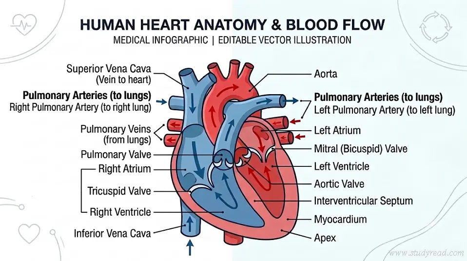
Cardiac Anatomy Quiz Questions
Each question also includes a full rationale to help you understand what you got wrong.
#1. 1. A 68-year-old patient presents with a mid-systolic crescendo-decrescendo murmur heard best at the right second intercostal space with radiation to the carotid arteries. Which anatomical structure is affected?
The aortic valve is located between the left ventricle and the ascending aorta. The aortic area is best auscultated at the right second intercostal space at the sternal border. Stenosis here produces a classic crescendo-decrescendo systolic murmur radiating to the carotids.
#2. 2. During a cardiac procedure, a surgeon, cautioning against damaging the AV node, focuses attention near the apex of the Triangle of Koch. Which internal anatomical landmark helps define this region in the right atrium?
The Triangle of Koch in the right atrium is bordered by the tendon of Todaro, the septal leaflet of the tricuspid valve, and the orifice of the coronary sinus. The AV node sits near the apex of this triangle.
#3. 3. A 22-year-old athlete undergoes an echocardiogram that reveals a bicuspid semilunar valve guarding the vessel that originates from the left ventricle. Which vessel-and-valve combination is being described?
Aortic valve The aortic valve is a semilunar valve located at the outflow tract of the left ventricle into the ascending aorta. Normally tricuspid, a congenital bicuspid aortic valve is a common anatomical variation predisposed to early calcification and stenosis.
#4. 4. An electrocardiogram of a patient with an acute myocardial infarction shows ST-elevation in leads V1 to V4, indicating an anterior wall infarct. Which coronary artery, running in the anterior interventricular sulcus, is most likely occluded?
The left anterior descending (LAD) artery branches off the left main coronary artery and runs along the anterior interventricular sulcus on the external surface of the heart, supplying the anterior wall of the left ventricle and the anterior two-thirds of the interventricular septum.
#5. 5. An emergency resident performs a pericardiocentesis on a patient with cardiac tamponade. As the needle passes from superficial to deep to enter the pericardial cavity and drain fluid, which layer is pierced immediately BEFORE reaching the pericardial space?
The pericardial cavity is a potential space situated between the parietal layer and the visceral layer (epicardium) of the serous pericardium. The parietal layer lines the internal surface of the outer fibrous pericardium.
#6. 6. Which chamber of the heart has the thickest myocardial wall?
The left ventricle generates systemic arterial pressure to perfuse the entire body, requiring significantly more muscular force than the right ventricle (which only pumps to the low-pressure pulmonary circuit). Its wall is typically 3x thicker.
#7. 7. A patient’s ECG shows a consistently prolonged PR interval with no dropped beats (first-degree AV block). Delay is most likely occurring at which structure?
The AV node normally imposes a brief physiological delay to allow ventricular filling before contraction. Pathological prolongation of this delay produces first-degree AV block, reflected as a prolonged PR interval on ECG.
#8. 8. A patient presents with an inferior wall MI, confirmed by ST-elevation in leads II, III, and aVF. Which coronary artery is most commonly responsible?
In most individuals (right-dominant circulation), the right coronary artery supplies the inferior wall of the left ventricle via its posterior descending branch, making it the most common culprit vessel in inferior MIs.
#9. 9. Following an inferior MI, a patient develops acute severe mitral regurgitation. Rupture of which structure is the most likely cause?
The posteromedial papillary muscle receives blood supply from a single source (typically the posterior descending artery), making it more vulnerable to ischemic rupture than the anterolateral papillary muscle, which usually has dual blood supply.
#10. 10. A newborn is found to have a continuous machine-like murmur heard throughout systole and diastole. This is most consistent with failure of closure of which structure?
The ductus arteriosus normally closes shortly after birth, becoming the ligamentum arteriosum. Patent ductus arteriosus (PDA) creates a continuous shunt between the aorta and pulmonary artery, producing the classic continuous murmur.
#11. 11. Which layer of the heart wall is composed of cardiac muscle tissue responsible for contraction?
The myocardium is the middle, muscular layer of the heart wall, composed of cardiac muscle fibers responsible for the heart’s contractile function.
#12. 12. A cardiology fellow points out a smooth vertical ridge on the internal posterolateral wall of the right atrium that separates the smooth-walled sinus venarum from the rough, muscular anterior atrium. What is this anatomical landmark called?
The crista terminalis is a smooth muscular ridge inside the right atrium that marks the junction between the embryological sinus venosus (smooth wall) and the atrium proper (rough wall with pectinate muscles). Externally, this corresponds to the sulcus terminalis.
#13. 13. During ventricular systole, which structures actively contract to pull on the chordae tendineae and prevent the eversion (prolapse) of the atrioventricular valves into the atria?
Papillary muscles project from the ventricular walls and attach to the cusps of the AV valves (tricuspid and mitral) via chordae tendineae. They contract during systole to maintain tension on the valves, preventing backflow and prolapse into the atria.
#14. 14. A patient is diagnosed with pulmonary regurgitation. Which valve structure prevents the backflow of blood from the pulmonary trunk into the right ventricle during ventricular diastole?
The pulmonary valve is a semilunar, avascular valve located at the base of the pulmonary artery. It consists of three semilunar cusps (designated as anterior, right, and left). Unlike AV valves, semilunar valves do not have chordae tendineae or papillary muscles.
#15. 15. An anatomical study highlights the base of the heart, which is formed predominantly by one specific chamber and lies directly anterior to the esophagus and thoracic aorta. Which chamber forms the majority of the heart’s posterior surface?
The base of the heart is located posteriorly and is formed predominantly by the left atrium (and to a lesser extent, the right atrium). Because the left atrium lies directly anterior to the mid-esophagus, transesophageal echocardiography (TEE) provides high-resolution images of this structure.
#16. 16. During a dissection of the right ventricle, students identify a distinct muscular bundle traversing from the inferior interventricular septum to the base of the anterior papillary muscle. What is this structure, and what critical element does it carry?
Right bundle branch The moderator band (septomarginal trabecula) is a prominent muscular band found in the right ventricle. It functions as a shortcut for the conduction system, carrying part of the right bundle branch of the AV bundle directly to the anterior papillary muscle for coordinated contraction.
#17. 17. A patient undergoing cardiac catheterization requires access to the main venous channel draining deoxygenated blood from the myocardium. Where does the coronary sinus reside externally, and into which chamber does it directly empty?
Right atrium The coronary sinus lies in the posterior part of the coronary sulcus (atrioventricular groove) on the diaphragmatic surface of the heart. It receives blood from the cardiac veins and empties directly into the right atrium, between the IVC opening and the AV orifice.
#18. 18. The ostia (openings) of the right and left coronary arteries originate from the aortic root immediately superior to two of the aortic valve cusps. Within which anatomical space are these openings located?
The aortic sinuses (Sinuses of Valsalva) are dilation spaces in the wall of the ascending aorta just superior to the aortic valve cusps. The right coronary artery arises from the anterior aortic sinus, and the left coronary artery arises from the left posterior aortic sinus. The right posterior sinus is non-coronary.
#19. 19. A standard frontal chest X-ray reveals an enlarged right heart border. Anatomically, which cardiac chamber or vessel forms the majority of the right border of the cardiac silhouette on a PA chest radiograph?
On a standard posteroanterior (PA) chest radiograph, the right border of the heart silhouette is formed primarily by the right atrium (bounded above by the SVC). The left border is formed mainly by the left ventricle and left auricle/aortic arch. The right ventricle forms the anterior (sternal) border and is not prominent on the right margin.
#20. 20. In an adult patient, an echocardiogram detects a probe-patent remnant of the fetal shunt that allowed blood to bypass the non-functional fetal lungs. Where is this landmark situated on the interatrial septum?
The fossa ovalis is a shallow, oval depression in the right atrial side of the interatrial septum. It represents the remnant of the fetal foramen ovale and its valve (septum secundum and septum primum), which allowed oxygenated placental blood to flow directly from the right atrium to the left atrium.
Results


Key Concepts Reviewed
- Crista terminalis — smooth ridge marking the junction between the sinus venosus and atrium proper in the right atrium
- Triangle of Koch — an anatomical landmark used to locate the AV node during procedures
- Moderator band (septomarginal trabecula) — carries the right bundle branch to the anterior papillary muscle
- Sinuses of Valsalva — aortic root dilations where the coronary arteries originate
- Fossa ovalis — adult remnant of the fetal foramen ovale
- Papillary muscles & chordae tendineae — prevent AV valve prolapse during systole
- Coronary artery territories — LAD (anterior wall), RCA (inferior wall), and their ECG correlations
If any of these felt unfamiliar, that’s normal — these are exactly the structures that separate a surface-level anatomy review from board-ready mastery.
Go Deeper
For a full video walkthrough of cardiac anatomy, conduction, and the clinical correlations covered in this quiz, Lecturio’s cardiovascular course series is a strong next step.
Keep Practicing
- Structure of Heart | Its Chambers, Valves, and Functions
- Digibind / DigiFab Dose Calculator (Digoxin Toxicity Antidote Protocol)
- Vancomycin AUC calculator
References & Recommended Reading
- National Center for Biotechnology Information (NCBI): StatPearls – Physiology, Coronary Circulation & Myocardial Perfusion. NLM / NIH Reference
- National Center for Biotechnology Information (NCBI): StatPearls – Physiology, Cardiovascular Hemodynamics & Cardiac Cycle. NLM / NIH Reference
- National Center for Biotechnology Information (NCBI): StatPearls – Physiology, Heart Sounds & Valvular Murmurs. NLM / NIH Reference
- University of Utah Health (Eccles Health Sciences Library): HyperHeart Interactive Cardiac Cycle & Electrocardiography Learning Center. University of Utah MedEd
- Yale School of Medicine: Clinical Cardiovascular Anatomy & Valve Mechanics Series. Yale MedEd
