The eye is the major sense organ dedicated to sight. It perceives the light energy and converts it into electrical energy, passing it to the brain so as to help us see the world.
The anatomy and physiology of the eye are quite simple but highly organized.
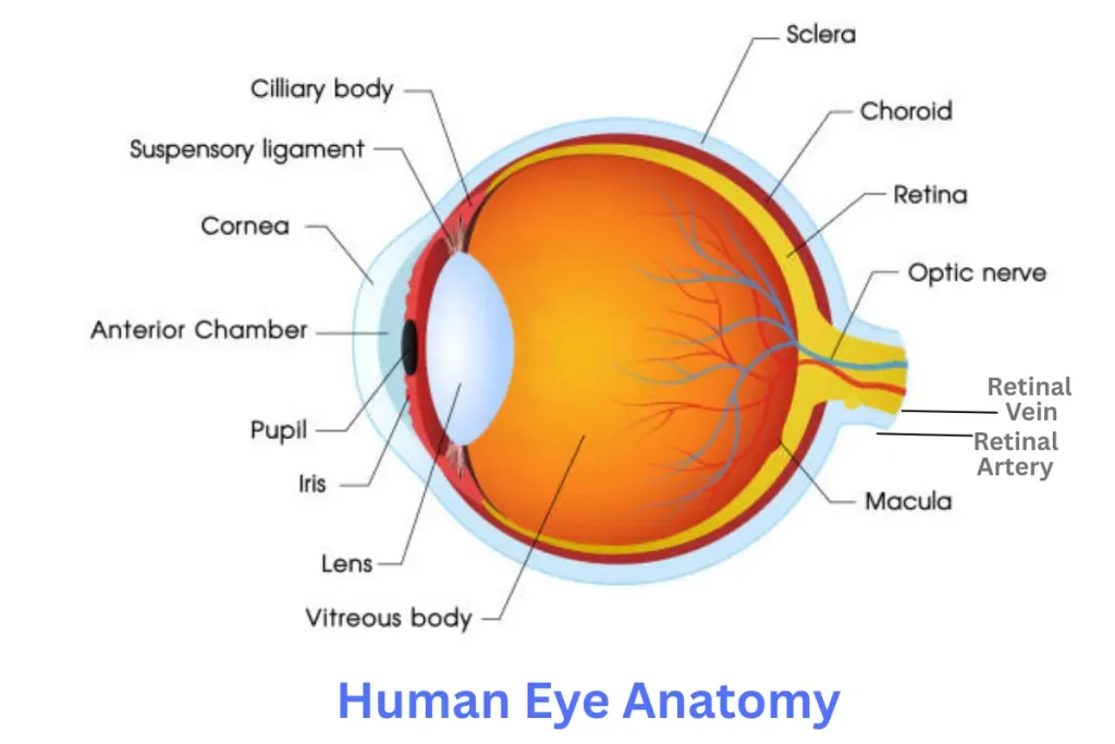
Human Eye Anatomy
Eyes are spherical-shaped organs fitted into the two orbits of the skull.
There are three layers in the eye, like
- The Outer layer: Sclera, cornea
- The Middle layer: Choroid, Iris, Ciliary body, and lens
- The Inner layer: Retina
The Outer Protective Layer
- It forms the outermost cover of eye anatomy.
- It is made of a dense, strong fibrous tissue that imparts shape to the eye.
- There are two distinct portions in this outer layer, as seen in the image above like.
- Posterior Sclera
- Anterior Cornea
Sclera:
- This is present in the posterior of the eye and forms 5/6th of the protective outermost covering of the eye.
- The gap between the sclera and the orbitals of the skull is filled with adipose tissue.
Cornea:
- This is a transparent portion of the outer layer that forms the anterior 1/6th of the eye.
- This cornea helps the light rays pass into the inner portions of the eyes.
- The junction of the cornea and sclera is called the limbus.
Functions of the outer protective layer.
- It helps fix the eyes in the orbitals.
- It protects the eyes from external damage.
- It gives shape to the eye.
The Middle Layer
- This is the second layer of the eyes that lies just below the outer layer.
This middle layer has the following parts
- Choroid
- Iris
- Ciliary body,
- Lens
Choroid
- It consists of a dense, capillary-rich layer that supplies blood to the eye.
- This choroid layer is dark brownish in color.
- It acts as a black screen, which prevents extra reflections inside the eyeball to get a perfect image.
Iris
- This is the colored part of the eye and is the extension of the choroid layer towards the anterior side of the eye
- This iris is pigmented, and can be dark, brown, green, blue, etc, based on genetics.
- There is a small orifice at the center called the pupil.
- Both parasympathetic and sympathetic nerves supply this iris.
- It dilates due to sympathetic stimulation for distant vision.
- It constricts by parasympathetic stimulation for short vision.
- This iris separates the aqueous chamber into anterior and posterior chambers.
Ciliary Body
- It is the continuation of the choroid layer anteriorly and has the ciliary muscle.
- From this ciliary body, the lens is suspended by suspensory ligaments.
- The epithelial cells in this ciliary body secrete the aqueous fluid.
Lens
- The lens is a biconvex, transparent, and elastic structure that is suspended by suspensory ligaments from the ciliary body.
- It helps to focus the objects based on their distance and concentrate the refracted light onto the retina.
- When it constricts, it becomes thicker, and when it dilates, it becomes thinner.
Inner layer (Retina)
- This is the innermost layer of the eye, made of nerve cells.
- This neural coat is an essential part of the eye for sight.
- It perceives the light and helps convert it into electrical energy.
- It has photoreceptor cells, namely the rods and cones.
- The rods are light-sensitive and recognize both dark and bright light.
- The cones are color sensitive and recognize the colors.
- The light received from the lens is converted into a nerve impulse and carried backward as the optic nerve into the brain for further processing.
- The point at which the nerves from the retina converge is called the optic spot.
- The retinal chamber is filled with vitreous humor.
Accessory parts of the eye
These are the parts that help in the safety and smooth functioning of the eye. They are
- The eyebrows
- The eyelids and eyelashes
- The lachrymal bodies
Eyebrows
- These are arch-shaped ridges present on the frontal bone.
- They have hair that is bent to one side.
- They help to prevent sweat, dust, and other waste from falling into the eye.
Eyelids and lashes
- Eyelids are delicate layers of tissue that cover the eye.
- They protect the eyes from dust, insects, etc., by their reflective action.
- The eyelashes are hair-like outgrowths projecting from the eyelids. They protect the eyes from insects and dust particles.
Lachrymal bodies
These are the glands that secrete tears.
These tears are rich in salts, lysozyme, and immunoglobulins. They help
a) Protection from irritants and dust.
b) Prevent microbial growth
c) Keep the eye moist, and the oil in it slows the evaporation.
Different parameters of the eyeball are given below:
- Anteroposterior diameter – 24mm
- Horizontal diameter – 23.5mm
- Vertical diameter – 23mm
- Circumference – 75mm
- Volume- 6.5ml
- Weight-7g
Segments and Chambers of Eyeball: Eyeball is divided into two segments, the anterior one and the posterior one.
- Anterior segment: The segmentation inside the eye is based on the position of the lens. An anterior segment consists of a crystalline lens hanging from the ciliary body by zonules and all structures in front of it, viz., iris, cornea, and two aqueous humor-filled spaces, i.e., anterior and posterior chambers.
- Anterior Chamber: The boundaries of the anterior chamber are anteriorly the back of the cornea and posteriorly the anterior surface of the iris and part of the ciliary body. It is 2.5mm deep in adults but varies in hypermetropes and myopes. It communicates with the posterior chamber, located just behind it, via the pupil, i.e., opening through the iris.
- Posterior Chamber: It is a small triangular structure located just behind the anterior chamber, guarded by the posterior surface of the iris and parts of the ciliary body anteriorly and by the crystalline lens and its zonules posteriorly.
- Posterior Segment: It consists of all structures that are posterior to the lens, i.e., vitreous humor, retina, and choroid, optic disc.
Extraocular Muscles: Extraocular muscles are responsible for the continuous movement of the eyeball to increase the field of vision. They can be divided into two groups of muscles:
- The Recti: There are four recti muscles :
- Superior Rectus
- Inferior Rectus
- Medial Rectus
- Lateral Rectus
- The Oblique: There are 2 oblique muscles :
- Superior Oblique
- Inferior Oblique.
Physiology of Vision: The physiology of Vision consists of three main mechanisms, they are:
- Initiation of vision (phototransduction), which is a function of photoreceptors, i.e., Rods and cones
- Processing and transmission of visual sensation is a function of image-processing cells of the retina and the visual pathway.
- Visual perception is a function of the visual cortex and related areas of the cerebral cortex.
Layers of the retina: The retina is the primary light-perceiving area of the eyeball, covering the whole inner surface of the eyeball. They are:
- The inner limiting membrane (ILM);
- the nerve fiber layer (NFL);
- the ganglion cell layer (GCL);
- the inner plexiform layer (IPL);
- the inner nuclear layer (INL);
- the outer plexiform layer (OPL);
- the outer nuclear layer (ONL);
- the outer limiting membrane (OLM);
- the photoreceptor layer (PL),
- The retinal pigmented epithelium (RPE) monolayer.
All these layers have individual functions in the perception of light.
- Phototransduction: The rods and cones are the cells that are designed to cause chemical changes when light falls on them. They serve as sensory nerve endings for visual sensation. When the light falls on them, a cascade of visual reactions starts, resulting in the generation of electrical changes. Rods contain a pigment called Rhodopsin or Visual Purple, while cones contain Iodopsin. Rods are responsible for dim light vision or monochromatic vision, while cones are responsible for color vision.
- Photochemical changes: The Photochemical changes include
- Rhodopsin bleaching
- Rhodopsin regeneration
Details about this are not discussed. But a picture is given to help find how NAD is produced, which thereby helps in electricity generation.
- Processing and Transmission of Visual Impulse
A receptor potential is developed in the photoreceptors, which is transmitted by electronic conduction, i.e., direct flow of current, not as an action potential, to other cells such as amacrine cells, horizontal cells, and ganglion cells. Now, these ganglion cells transmit electric impulses to the action potential to neurons of the lateral geniculate bodies and later to the primary cortex.
- Visual perception
It consists of some thresholds, for example:
- Light sense: It refers to the minimal brightness required to evoke a sensation of light, called the light minimum. It should be measured after at least a dark adaptation of 20 to 30 minutes. The two types of visual adaptations are
- Light adaptation
- Dark adaptation
Rods are more sensitive to low light vision, i.e., scotopic vision. Therefore, rods are used more in dim light vision, whereas cones are used more in bright light vision, i.e., photopic vision.
The dark adaptation depends on the presence of vitamin A. So, a deficiency of vitamin A causes night blindness.
The eye is the only organ of photoreception. It lets us see the world. Many diseases affect the eye, ranging from minor ailments like hypermetropia, myopia, and cataracts to diseases of significant concern like glaucoma, retinopathy, etc.
Also, see cow eyes anatomy.

Thanks for the feedback. 🙂
Very well explained -it kept me focussed until the end which is not always the case in explanations.