Skip to content
Menu
Pharmacology
Anatomy
Biology
Microbiology
Biotechnology
Chemistry
Pharma Analysis
Resources
🧮 Medical Calculators
Education
Study skills
Education technology
Physics
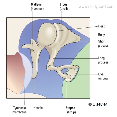
ear ossicles
Leave a Comment
Comment
Name
Email
Website
Save my name, email, and website in this browser for the next time I comment.