The roots are a very important organ in plants. The root anatomy is different for monocot and dicot plants.
Roots develop from the radicle and help in the absorption of water and minerals from the soil that are required for plant life.
The roots fix the plant to the ground and provide support to the part of the plant above ground. In a few plants like rhizomes, roots also help in storing the reserve food produced by photosynthesis. Thus, it is important to know the internal root.
The Anatomy of Root
The root’s anatomy is different for monocot and dicot plants. Hence, we will see them separately as
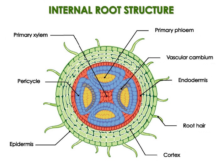
The Anatomy of Dicot Root
The primary dicot root appears circular in transverse sections and cylindrical in outline. The tissue layers present in dicot root from outside to inside are
- Epiblema,
- Cortex,
- Endodermis,
- Pericycle, and
- Vascular strands.
If there is a presence of pericycle and pith, they are considered to be a part of the stele along with vascular bundles.
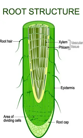
Epiblema
It is the outermost layer in a young root and is devoid of stomata and cuticle.
The cells have thin walls and are tubular in shape. Some of the epidermal cells are extended to form root hairs, which are thin-walled tubular structures.
These modified cells are called root hair cells or trichoblasts.
Epiblema, also called rhizodermis, means the skin of the root. Root hairs have an external pectose layer, which helps them push through the soil interspaces to absorb water and mineral salts. They have a lifespan of seven days, after which they shrivel up.
Cortex
This contains several layers of thin-walled parenchyma cells, which may be rounded, oval, or angular in shape. These cells store food. Intercellular spaces are less in volume but abundant in number. The cortex conducts water and minerals from the root hair to the interior of the root.
Endodermis
The endodermis is the modified inner layer of the cortex. The cells appear barrel-shaped in transverse sections with no intercellular spaces. These cells have lignified and suberised bands of thickening called Casparian strips. Casparian strips do not allow movements of conducting material to and fro the cortex in the intercellular spaces (apoplastic pathway). In this layer, the conducting materials have to enter inside the cell(symplastic pathway) for transport to the inner layers. These Casparian strips are present in both radial and tangential walls.
Pericycle
Made up of parenchymous cells, it is the outer boundary of a stele or a vascular strand. It may be single (sunflower) or multiple (mulberry) layered. The pericycle is significant in the root because it is the place of formation of root branches. It also gives rise to cork cambium or phellogen and a small part of the vascular cambium.
Vascular strand
This consists of 2 to 6 radial bundles, each of xylem and phloem, which lie on different radii and in alternate locations. Xylem bundles are large and wedge-shaped with differentiated protoxylem. Xylem is exarch. Phloem bundles are small and oval and remain in contact with the pericycle. The protophloem is undifferentiated. Conjunctive parenchyma tissue present between the xylem and phloem bundles separates the channels and produces vascular cambium. The phloem consists of companion cells and phloem parenchyma. The presence of fibers is rare. The xylem consists of vessels and a few tracheids.
Pith
It is mostly absent if present; it is quite insignificant. It is made of parenchyma cells that store food and waste products.
Anatomy of monocot root
Normal secondary growth is absent in the monocot root. The layers in monocot root from outside to inside are
- epiblema,
- cortex,
- exodermis,
- endodermis,
- pericycle,
- vascular stand,
- pith.
Epiblema
An outermost, single layer of thin-walled cells that do not have intercellular spaces, cutin, or cuticle. Some of the cells form outgrowths called root hairs, which are covered by a gummy pectin layer, which helps them push to soil interspaces for the absorption of various materials.
Cortex
Consists of multiple layers of thin-walled oval or spherical parenchymal cells with intercellular spaces. These cells store food and also help in the radial movement of water and minerals from the epiblema to the interior. The epiblema ages and shrivels up, becoming suberised and thick-walled. This forms the exodermis. In some cases, sclerenchyma also develops inner to exodermis, providing extra support.
Endodermis
The inner boundary of the cortex covers the inner stele. It consists of a single layer of barrel-shaped cells with no intercellular spaces. The cells possess suberin and lignin thickenings called Casparian strips occurring over radial and tangential walls. This thickening regulates the passage of substances through it. The cells have plasmodesmata to allow the passage of substances.
The stele consists of pericycle vascular bundles and pith.
Pericycle
It is the outermost layer of the stele that lies below the endodermis. It may be single-layered (maize) or multi-layered.
It consists of parenchymatous cells. In older roots, the cells are thick-walled. Root branches develop from the pericycle.
In monocot roots, the pericycle does not form a cambium because there is no secondary growth in monocots.
Vascular strand
It consists of eight or more radial bundles, each of the phloem and xylem arranged alternatively. It is polyarch. The number of xylem and phloem bundles is equal. They are arranged in contact with the pericycle and are alternate to each other.
Phloem patches are present in the form of small oval bundles made up of sieve tubes and companion cells.
The presence of phloem fibers is rare, and phloem parenchyma is absent. Protoxylem and protophloem remain in contact with the pericycle. Metaxylem is towards the inner side.
The xylem consists of vessels and parenchyma with few tracheids and fibers. Metaxylem is large and fewer with pitted thickening.
Protoxylem vessels are narrower and smaller, having annular or spiral thickenings. The conjunctive tissue layer is present between phloem and xylem bundles.
In younger plants, the conjunctive layer is parenchymatous (stores food and helps in conduction) and later becomes sclerenchymatous (provides mechanical strength). It does not form vascular cambium.
Pith
It is present in the center of the root and consists of parenchymatous cells. The cells may be rounded or oval in outline with small intercellular spaces. The cells store food.
The pith cells may be thick-walled or sclerified to provide support in monocots growing in drier areas.
Also, see storage roots.
