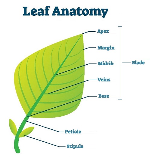
Leaves are the main photosynthetic organs of the plant. Anatomy of the leaf is the detailed study of the internal structure of a leaf, usually revealed by its dissection. Leaves are responsible for converting sunlight and carbon dioxide into glucose, which is used to provide energy to the plant.
 Leaves are classified into mainly two types based on their structure, dorsiventral and isobilateral. There is another type of leaf called unifacial, like Allium. A unifacial leaf is cylindrical in outline, so there is no distinction between the upper and lower surface. In all leaves, there is no distinction between protophloem (older phloem) and metaphloem (new phloem).
A dorsiventral leaf is also called a bifacial leaf and is present mostly in Dicot plants. Isobilateral leaves are further classified into two types-
- with undifferentiated mesophyll
- with differentiated mesophyll
Dorsiventral Leaves
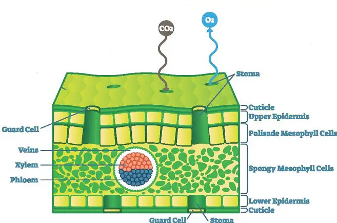
The dorsiventral leaf is flattened with two distinct surfaces. The upper surface is darker and is called the adaxial surface. The adaxial surface faces the sun. The lower surface is lighter and is called the abaxial surface. This faces downwards. The abaxial surface may also be called the dorsal surface. The veins can easily be seen over the surface. The dicot leaf is divided into the upper epidermis, lower epidermis, mesophyll, midrib, and vascular strands.
Upper epidermis
The covering present over the upper surface of the leaves is called the upper epidermis. The upper epidermis may occur in single or more layers. The cells are compactly arranged to minimize intercellular spaces for more protection. The cells do not have chloroplasts and are transparent to transmit light rays. The internal cells the epidermal cells are rectangular or barrel-shaped. For extra protection, the outer walls are thicker compared to the inner walls and a layer of the cuticle is present on the outside. Stomata are present in mesophytic forms and if present in other forms there are less in number. In some cases, the leaves have hair.
Lower epidermis
It is the covering over the lower surface of the leaves. It is usually single layered. These cells are rectangular or oval in shape. Due to the absence of chloroplast, the cells are transparent. The outer walls are thickened and contain cutin. The outer side also has a layer of cuticle for extra protection and reducing transpiration rate. Hair may also be present on the lower epidermis. The lower surface contains a large number of stomata. These stomata have kidney-shaped guard cells that contain chloroplast. Specialized epidermal cells called subsidiary cells surround the guard cells. The guard cells may be at the level or sunken below the level of the epidermis.
Mesophyll
Formed mainly of chlorenchyma cells, this is the ground tissue of the leaf and is the main photosynthetic region. The mesophyll is a dorsiventral leaf that is divided into upper palisade and lower spongy. The palisade parenchyma lies in contact with the upper epidermis and the cells are columnar in shape. Palisade is richer in chloroplast and their full most photosynthetic activity takes place here.
Spongy parenchyma lies below the palisade parenchyma, and the cells are variable in shape with thin cell walls. Large intercellular spaces are present, which are in contact with the stomata through substomatal cavities. Fewer chloroplasts are present, and therefore it is lighter in color.
Vascular strand
This constitutes the vascular bundles present in the mesophyll. Each vascular bundle is encircled with a layer of thick-walled bundle sheath. The cells are compactly arranged and do not possess chlorophyll. Within the vascular bundle, xylem is present on the adaxial side, and the phloem is present on the abaxial side. Sclerenchyma fibers may be present within the bundle sheath. Large-sized sclereids called idioblasts may also be present. Intrafascicular cambium is absent, so the vascular bundles are closed. Vascular bundles are conjoint and collateral with no distinction between protoxylem and metaxylem.
Midrib
On the upper surface, there is a depression where the midrib or larger veins are present, while on the lower side, there is a prominent ridge. The mesophyll is absent in these regions, but collenchyma or sclerenchyma is present. The hypodermis layer also is present. These ribs provide mechanical strength to the leaf and also help in the transfer of substances. They are surrounded by parenchymatous ground tissue.
Isobilateral leaves
Usually present in monocots, both surfaces are equally green, and either side may face the sun. The components of the leaf are the epidermis, mesophyll,l vascular strand, and midrib.
Epidermis
Present on both surfaces, this is the outermost covering of the leaf. The cells are parenchymatous and rectangular in shape. They are transparent and do not have chloroplast. The surface may also have cutin and silica deposits for protection. The cuticle is present to minimize the transpiration rate. In some plants, the adaxial epidermis has large, colorless, thin-walled bulliform cells. When water is deficient, the cells become flaccid, making the leaves curl inwards to lessen the surface area, thus minimizing water loss. When water levels become normal, the bulliform cells become turgid; the leaves become flat.
Equal numbers of stomata are present on both surfaces. The guard cells are dumbbell-shaped and the stomata may or may not have subsidiary cells. Each stoma leads into a substomatal cavity.
Mesophyll (ground tissue)
The mesophyll of isobilateral monocot leaves is undifferentiated. The cells are all similar, oval or round-shaped. Small intercellular spaces are present, which are connected to substomatal cavities. The cells are parenchymatous and contain chloroplast. Photosynthesis can thus take place.
Vascular strand
Smaller vascular bundles lie in the middle of the mesophyll, while the larger ones occupy the whole area between the surfaces. The bundle sheath and its extensions are sclerenchymatous. The bundles are covered by chlorenchyma cells. The sheath may be single or double-layered. The vascular bundles are conjoint, collateral and closed. The phloem is present towards the abaxial side and the xylem towards the adaxial side. In smaller vessels, there is no distinction between protoxylem and metaxylem. In larger vessels, they are distinct, and the protoxylem cavity may be present. Metaxylem vessels have pitted thickening civil protoxylem has annular or spiral thickening.
In some plants, the mesophyll forms concentric layers around the vascular bundles such that the chloroplasts are arranged centrifugally around the bundle sheath cells. This is called Kranz anatomy, and it increases the efficiency of photosynthesis. It is more common in tropical conditions and may also be found in some dicot leaves.
Midrib
It is the thickest part of the leaf. Other large veins may also have similar thickenings. In the upper adaxial surface, a shallow groove is present while a ridge is present in the abaxial surface in the midrib area. The mesophyll is absent, and there are sclerenchymatous and parenchymatous patches on both sides.
