A microscope is a device used to visualize objects invisible to the naked eyes. These instruments magnify the image many times to be visible to us.
The invention of the microscope has revolutionized science, medicine and other areas where the microscopic structural examination is required. Microorganisms like the bacteria, fungi, protozoans, etc. were found to be the actual causes of epidemic diseases.
Types of microscope available
- Simple microscope
- Stereoscopic microscope
- Compound microscope
- Phase contrast microscope
- Electron microscope
With the advancement of technology, one can even get an image of the virus and minute molecular structures. Besides, it is possible to take a photograph of tissues and also label the specimens pictures by connecting them to computers. Let us see them in detail.
Simple microscope
This is a just microscope with a single magnification lens like 10x.
It is used to visualize any small objects which are so minute and not visible to the naked eye.
Stereoscopic dissecting microscope
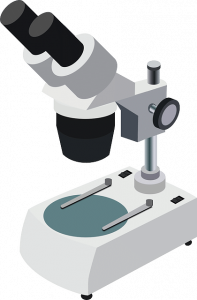
This microscope is commonly used to study and to identify the opaque objects. It is generally used for low power observation, dissection, etc. Its magnifying power varies from 4x to 40x or even 60x. It has two objectives and two eyepieces which help to make a 3D image of an object.
Compound microscope
A Compound microscope is the most commonly used microscope used in labs performing biological experiments and clinical diagnosis. 
It has an eyepiece and 2 or 3 objectives lenses. The magnification power ranges from 5x to 8x, 10x to 15 x with the magnification of the objectives that range between 10x or 40x. It is used to examine semitransparent and translucent objects or part of objects in the form of thin slices. Most bacteria, protozoa, blood cells can be observed by using it.
Phase contrast microscope
Phase contrast microscope is commonly used to study biological tissues and specimens. It is also known as one of the most important tools in medical research. It is generally used to study the specimens that are colorless and transparent and usually difficult to distinguish from their surroundings. These specimens or objects are commonly termed as phase objects. All phase contrast microscope is not the same but, they work on the same principle and have five eyepieces of different magnification that includes 10x, 20x, 40x, 100x and Bright Field (BF).
The electron microscope
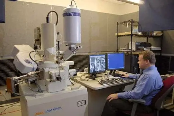
This is the ultimate microscope to study any know minute particle. It is used to study a variety of biological and non-biological products such as the structure of cell organelles, DNA, RNA, virus, biopsy sample, metals, crystals, and large molecules. It is one of the important tools for biological and medical sciences as it magnifies the image of an object by 10,000,000x unlike other types, where one does not see the object under study with a naked eye. The instrument produces images of the objects under study and one needs to observe the images. In the electron microscope, two types of lenses are present like “electrostatic” and “electromagnetic.” These types help to control the beam of the electron which helps to illuminate the specimen and produces its magnified image.

The electron microscope is of five different types:
1. Transmission electron microscope (TEM)
2. Scanning electron microscope
3. Reflection electron microscope
4. Scanning transmission electron microscope
5. Low-voltage electron microscope.
The electron microscope is very expensive and requires a separate chamber for its housing. Besides one needs to have a nitrogen plant for use during operation as it generates a large amount of heat.
Currently, microscope finds their use in biology, medicine and even research.