The anatomy of a cockroach can be viewed as the external and the internal anatomy. It is an insect and its structure is similar to most other insects.
Cockroach Anatomy and Structure
The anatomy has two main parts like the
1) External
2) Internal
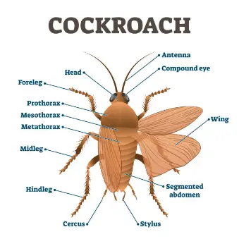
External anatomy includes the
- Appendages
- eyes
- antenna
- cercus
- stylus
- wings
- prothrax
- mesothorax and
- metathorax.
The internal anatomy consists of the
- Body wall and exoskeleton
- Coelom and body cavity.
Body wall and exoskeleton
The body wall (integument) consists of a
- cuticle,
- hypodermis, and
- basement membrane.
- The entire body of the cockroach, including the appendages, is covered with a thick, brown-colored, non-living, hard, chitinous substance called the cuticle.
- This cuticle forms the exoskeleton of the insect and is secreted by the hypodermis found underneath.
- The cuticle is made up of chitin, a horny protein. It is chemically an amino acid polysaccharide that is water-insoluble.
- The cuticle is hard due to the sclerotization and the resultant hardened plates are called sclerites.
- At the joints (sutures), sclerites are connected by a soft, flexible and arthrodial membrane that allows movements of the body and appendages.
- In each thoracic and abdominal segment, there are 4 sclerites found, which include a dorsal tergum, a ventral sternum, and two very small lateral pleura.
- The cuticle also lines the foregut, hindgut, trachea, and genital ducts of cockroaches.
- Several invaginations of the cuticle are there, which is called tentorium, which is present in the form of an endoskeleton in the head region.
- The thorax side endoskeleton is called apodemes.
- The hypodermis is located beneath the cuticle, which it secretes.
- The hypodermis is made up of columnar cells, which are highly organized in a single layer.
- All the cells of the hypodermis rest on the basement membrane and are anchored to it by hemidesmosomes.
- Basement membrane bounds the inner surface of the hypodermis.
- The basement membrane consists of an amorphous granular material, which is most probably a mucopolysaccharide.
Coelom and body cavity
In cockroaches, the alimentary canal and other viscera are enclosed in the hemocoel, which contains blood.
The true coelom in cockroaches is found in a much-reduced state and is found only in the cavities of reproductive organs only.
Digestive system:
The digestive system in cockroaches is the most conspicuous organ system of the body.
The system includes the mouthparts and alimentary canal.
The alimentary canal is long and coiled up with varying diameters.
It can be divided into three parts like
- foregut
- midgut and
- hindgut.
- The foregut and hindgut are lined with ectodermal cells, which are secreted by the cuticle of the ectoderm.
- At the same time, the midgut is lined with mesodermal cells.
- The midgut lacks the cuticular lining and is capable of absorbing digested food.
- The foregut is also called the stomodeum, which includes the mouth cavity, pharynx, esophagus, crop and gizzard.
- Food is crushed in the mouth cavity and well mixed with saliva and leads to the pharynx, which is a tubular and short structure folded posteriorly.
- From the pharynx, the esophagus arises, which is a straight narrow and laterally compressed tube.
- The crop is a large, thin-walled, pear-shaped sac that extends up to the fourth segment of the cockroach’s body.
- The crop is the largest part of the foregut.
- The epithelial lining and cuticular lining are highly folded.
- A network of the trachea covers the outer structure of the crop. The crop is responsible for the storage of food.
- Crop leads to the gizzard, which is small, cone-shaped, muscular, and thick-walled.
- The gizzard is also called proventriculus, which marks the end of the foregut.
The gizzard comprises two parts
- An anterior armarium and
- A posterior stomodael valve.
- The armarium has 6 longitudinal folds that are internally reduced in the lumen.
- The stomodael valve is behind the posterior end of the gizzard, which extends into the lumen of the midgut.
- The midgut or mesenteron is a short, narrow tube-like structure that forms the middle part of the alimentary canal.
- The midgut is internally lined up by the glandular epithelium and forms the true stomach, which serves the function of digestion and absorption.
- From the junction of the midgut and hindgut arise 80-90 very narrow, thread-like yellow-colored blind tubules, which are called Malpighian tubules.
- These tubules are excretory in function.
- The posterior portion of the alimentary canal called the hindgut is divided into three regions: ileum, colon and rectum.
- The ileum is a short, narrow tube that is characterized by 6 tiny triangular lobes internally.
- The colon is longer and wider than its irregular shape.
- The rectum is an oval or spindle-shaped sac that has external ridges alternating with longitudinal thickenings called rectal pads.
- The rectum opens to the anus.
Circulatory system
- The cockroach has an open or lacunal circulatory system that flows freely within the body cavity or hemocoel.
- The blood in cockroaches is also called hemolymph.
- There are a heart and aorta and the capillaries for the pumping of blood.
- The body cavity is called the hemocoel, which is filled with blood .haemoceol is divided by a dorsal and a ventral diaphragm. the three sinuses are
1. dorsal pericardial
2. middle perivisceral and
3. Ventral perineural.
- Diaphragms have pores or fenestrae to permit hemolymph from one sinus to others.
- The heart is enclosed by the dorsal pericardial sinus, which lies mid-dorsally beneath the terga of the thorax and abdomen.
- It is a long, narrow tube with the anterior part open and the posterior part closed.
- It consists of 13 funnel-shaped chambers or segments, each communicating by the valvular opening with the one in front of it.
- The hinder end of each chamber has a pair of minute lateral openings, the Ostia.
- These allow the flow of the hemolymph from the pericardial sinus to the heart only and not in the reverse direction.
- The hemolymph of the cockroach comprises a clear, colorless plasma that is rich in amino acids, uric acids, and numerous different types of cells, called hemocytes.
- The hemolymph is devoid of any respiratory pigment and does not assist respiration.
Respiratory system
- The respiratory system of the cockroach is well-developed to compensate for the poorly developed circulatory system.
- The respiratory system consists of the trachea, tracheoles, and spiracles.
- The hemocoel consists of a network of elastic, closed, and branching air tubes which are called trachea.
- The trachea includes pairs of large, parallel, longitudinal tracheal tufts that are connected by transverse commissures.
- The trachea is formed by the invaginations of the outer integument; thus it is made up of epithelial tissue.
- The cuticular lining is gradually thickened, forming the initium or taenidia.
- The trachea profusely branches into smaller tubes called tracheoles, which anastomose and penetrate all over the body.
- The tracheoles have a diameter of 1 micron.
- They are impermeable to water.
- The main tracheal trunks open to the exterior on the body surface by small 10 pairs of segmentally arranged apertures called spiracles or stigmata.
Excretory system
- The main structures of the excretory system of cockroaches are malpighian tubules, fat body cells, uricose cells, and cuticles.
- The malpighian tubules are attached to the alimentary canal at the extreme anterior end of the hindgut.
- These tubules are fine, long, unbranched, yellowish, and blind lying freely in the hemolymph.
- The tubule is lined up by glandular epithelium, which has a characteristic brush border.
- A fat body is found in the greater part of the hemocoel, which is lobed and white tissue.
- The fat body cells have different types of cells, but only urate cells are associated with excretion.
- Uricose glands are the mushroom gland of the cockroach, which possesses long, blind tubules that are at the periphery.
- These tubules store uric acid and discharge it over the spermatophore during copulation.
Nervous system
- The nervous system includes the central, peripheral, and sympathetic or stomatogastric nervous systems.
- The brain and ventral nerve cord, with its ganglia, form the central nervous system.
- Brain or supra-oesophageal ganglion is a large and bilobed structure located above the head region.
- The three parts include protocerebrum, deutero-cerebrum and tritocerebrum.
- The remaining three pairs of ganglia of the head fuse to form the sub-oesophageal ganglion which lies below the esophagus.
- The ventral nerve cord is double-layered and posteriorly runs along the mid-ventral line of the thorax and abdomen.
- It bears 9 ganglia, 3 in the thorax, and 6 in the abdomen.
- The nerves are given off from the ganglia to all the parts of the body, which is called the peripheral nervous system.
- The protocerebrum of the brain gives off paired optic nerves to the eyes, deutero-cerebrum gives off paired antennary nerves to frons and labrum.
- The sympathetic nerves comprise ganglia and a retro-cerebral complex.
- The frontal ganglion lies above the pharynx in front of the brain; it sends nerves to the pharynx, clypeus and labrum.
- It is connected to the protocerebrum of the brain by a neuronal connective system.
