A male gamete (sperm) fuses with a female gamete (ovum or egg) to produce a zygote that develops into an individual. But humans are diploid while gametes are haploid.
So the cells undergo meiosis to produce haploid gametes. This process is called gametogenesis.
In men, it occurs as spermatogenesis, and in women, it occurs as oogenesis.

Spermatogenesis and oogenesis Steps
Spermatogenesis occurs in testes of man.
Spermatogenesis
The process of the development of male germ cells into spermatozoa is called spermatogenesis. The duration of spermatogenesis is 65-74 days. The primitive or immature germ cells called spermatogonia are present in the testis. The testes also contain supporting cells called Sertoli cells and testosterone-producing cells called Leydig (interstitial) cells. Spermatogonia are located attached to the basement membrane of the seminiferous tubule.
Spermatogenesis starts at puberty when under the influence of rising levels of gonadotropin and testosterone, the inactive germ cells are activated and spermatogenesis is initiated. Thereafter spermatogenesis continues throughout life. Spermatogenesis can be divided into three distinct phases-
- Mitosis
- Meiosis
- Spermiogenesis
Mitosis
The primitive germ cells present in the semeniferous tubules undergo repeated mitotic divisions to form primary spermatocytes. This process is called spermatocytogenesis. These mitotic divisions produce two types of spermatogonia A and spermatogonia B.
- Spermatogonia A- they resemble the original spermatogonia and are the source for the subsequent spermatogonia in the testis. They serve as a reserve and undergo further division to produce more spermatogonia.
- Spermatogonia B- they grow and enter into the add nominal component, where they develop into primary spermatocytes.
Meiosis
The primary spermatocytes are diploid and have to undergo two meiotic divisions to produce haploid cells.
The first meiotic division produces two secondary spermatocytes that are haploid.
The second meiotic division of these secondary spermatocytes produces haploid spermatids.
These spermatids contain 23 chromosomes, i.e., 22 autosomes and 1 sex chromosome.
Thus Each primary spermatocyte forms four spermatids, two of which contain an X chromosome and two of which contain the Y chromosome.
Each spermatogonium divides into 512 spermatids after these two stages.
Spermiogenesis
Spermiogenesis is the process of development of the haploid spermatids into mature spermatozoa.
Spermatids are small and round and have to undergo many structural changes to form spermatozoa. The changes it undergoes are
- A large reduction in the cytoplasm- cytoplasmic fragments are discarded in the form of residual bodies.
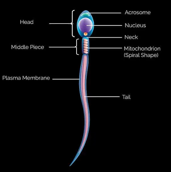
- The nucleus elongates to become the head of spermatozoa
- An acrosomal cap is formed, which contains enzymes needed to penetrate the ovum from over the head.
- The middle piece and tailpiece form so that the spermatozoa can move swiftly within the uterus.
These changes occurring in mature sperm help them survive in the acidic environment of the vagina and help them recognize, move towards, and fertilize the ovum.
Spermiation
Spermatozoa, after being formed, stick to the Sertoli cells in the lumen of seminiferous tubules.
The detachment of the head of spermatozoa and its release into the luminal fluid is called spermiation.
Capacitation
It is the functional maturation of sperm. This occurs in the female genital tract. The glycoprotein of the sperm cell membrane is eroded, leading to exposure of acronym receptors.
Motility increases and an acrosomal reaction occurs. The acrosomal reaction is where the sperm cell membrane fuses with the ova cell membrane during fertilization.
Oogenesis
The process of the development of female germ cells into spermatozoa is called oogenesis.
While spermatogenesis starts at puberty and continues throughout life, the process of oogenesis starts in fetal life and ceases at menopause.
The spermatogenesis process is short, while the development of each oocyte begins in intrauterine life and is completed at ovulation, which occurs during the menstrual cycle.
Many sperms can be produced within a few days, while a single ovum is produced during each cycle by follicle maturation.
In embryonic life, the primordial germ cells migrate from the yolk sac to the genital ridge in the 6th week of gestation. These oogonia undergo repeated mitotic divisions until the number reaches 7 million. After mitosis ceases, the oogonia are called oocytes.
Oocyte development occurs in three stages- oogonium becoming primary oocyte, primary oocyte converted to the secondary oocyte, and finally secondary oocyte developing to mature ovum.
Oogonium becomes the primary oocyte.
Oogonium converts to oocytes in embryonic life.
These diploid oocytes are called primary oocytes and they undergo two meiotic divisions.
The first meiotic division occurs in primary oocytes in intrauterine life in the 8th week of gestation.
The cells get arrested in a prophase state, but the oocytes grow in size.
Oocyte degeneration starts from intrauterine life such that only about 1 million primary oocytes remain at the time of birth.
By the time of puberty, only 200,000 remain in the ovary. In a woman’s life, only about 400 oocytes are ovulated and others degenerate.
Thus the process of oogenesis is limited as new oogonia cannot be manufactured in the ovary.
Primary oocyte converted to secondary oocyte
The primary oocyte that is destined for ovulation completes its first meiotic division just before ovulation.
This division results in two structures the larger daughter cell in the secondary oocyte containing 23 chromosomes and the other first polar body, which is smaller.
Thus the cytoplasmic division is grossly unequal and the polar body is completely non-functional.
Secondary oocyte forming ovum
Secondary oocyte undergoes second meiotic division after ovulation, but the process is arrested in metaphase.
The division is completed only when the sperm penetrates the egg.
Thus the egg or ovum contains 23 chromosomes and the second polar body forms during the second meiotic division.
Each primary oocyte produces only one ovum.

Ovarian Follicles
The oocyte grows throughout its life in the ovarian follicle till ovulation when the ovum is released from the follicle.
Along with the development of oocytes in ovarian follicles, follicles also grow in different phases.
Each primary oocyte gets surrounded by a layer of granulosa cells to form the primary follicle.
The primary follicles get surrounded by more layers of granulosa cells and new theca to form secondary follicles.
The tertiary follicle forms from the secondary follicle and has a characteristic fluid-filled antrum.
The theca layer is distinguished into inner theca internal and outer theca externa. In this stage, the primary oocyte completes its first meiotic division.
The tertiary follicle matures into the final Graafian follicle. A membrane called zona pellucida is formed around the secondary oocyte.
The Graafian follicle ruptures to release the ovum during ovulation.
Similarities between oogenesis and spermatogenesis
- Both take place in the reproductive organs of the body.
- Both the sperm and ova originate from the germinal epithelial cells.
- Both the process is controlled by anterior pituitary gland hormones.
- Both produce haploid gamete cells (1X) from the original diploid cell.
- Both undergo 2 meiotic divisions to divide chromosome numbers by half.
- In both cases, nuclear and cytoplasmic changes occur in the respective cells.
- In both cases, the first mitosis occurs to cause the multiplication of cells.
15 Differences between spermatogenesis and oogenesis.
| Sl.No | Feature | Spermatogenesis | Oogenesis |
|---|---|---|---|
| 1 | Occurs | Occurs in male | Occurs in female |
| 2 | Organs involved | Semeniferous tubules in the testis | Ovaries |
| 3 | Effecting Hormones | Testosterone and follicle-stimulating hormone (FSH) regulate the maturation process. | Estrogen, FSH, and luteinizing hormone regulate the maturation of ova. |
| 4 | Active age | In males, it activates from puberty and runs till old age or death. (14th year onwards) | In females’ it is active from birth and ends at menopause. (From birth to 50 years approximately) |
| 5 | The timespan of the process | It takes 64 days for sperm formation | It takes 10 days for the germinal cells to mature into ova and get released. |
| 6 | Temperature | Spermatogenesis occurs at a temperature less than 3 degrees from the body temperature. | Oogenesis occurs at normal body temperature. |
| 7 | Number of gametes | About 1500 sperms are made every second | Only one ova is released for fertilization. |
| 8 | Cytoplasmic division | Equal cytoplasmic divisions occur in germinal cells. | Unequal cytoplasmic divisions occur in germinal cells |
| 9 | Quantity of Food reserve | Less food reserve is present in sperm as it is surrounded by seminal fluid containing nutrition. | More food reserve is in ova. |
| 10 | Numbers per cell | Four motile male gametes i.e. sperms are formed from one | Only one nonmotile female gamete i.e. ovum is formed |
| 11 | Recycling time | Many sperms are formed within a few days | A single ovum is produced during each cycle (28 days) |
| 12 | Meiotic division status | The meiotic division is always completed | The meiotic division is not completed until sperm penetrates zona pellucida |
| 13 | Duration of the whole process | The duration of the spermatogenesis process is short | Development of each oocyte begins in intrauterine life and is completed at ovulation, so it has a long duration |
| 14 | Meiotic division specialty | Meiotic divisions are not arrested | Meiotic divisions are arrested |
| 15 | Numbers (capacity) | Unlimited process, no predetermined number | The process of oogenesis is limited to a fixed number of oocytes present in the ovary, more cannot be formed. |
