Bacterial identification is the process of determining the taxonomic identity of an unknown bacterial isolate through its physical, biochemical, and molecular characteristics.
It is essential for clinical diagnostics, pharmaceutical quality control, and epidemiological tracking,
| Stage | Method / Test | Key Objective | Examples |
| 1. Morphology & Staining | Gram Stain, Acid-Fast Stain | Determine cell wall type, shape, and arrangement | Gram-positive cocci in clusters (Staphylococcus) |
| 2. Culture Characteristics | Selective & Differential Media | Observe colony morphology, hemolysis, or sugar fermentation | Lactose fermentation on MacConkey agar |
| 3. Primary Biochemicals | Catalase, Oxidase, Coagulase | Differentiate major bacterial groups | Catalase (+) Staphylococcus vs. Catalase (-) Streptococcus |
| 4. Rapid / Automated | API Strips, VITEK | Multi-test biochemical profiling | Enteric pathogen identification |
| 5. Molecular / Modern | 16S rRNA, MALDI-TOF MS | Definitive genetic or protein-mass identification | Outbreak tracking & rare isolate ID |
Bacterial Identification Methods
So we identify them using a microscope, by increasing their population to a large enough number, or by biochemical methods.
Hence, in identifying bacteria, we use microscopical, medical, biochemical, & serological methods.

1. Identification by morphology
This method is used to determine the physical appearance of individual bacteria.
a. Based on size
- Here, bacteria are identified by their physical size, such as small or large, expressed in microns.
- For this, bacteria are viewed under a microscope to consider their size. One can apply a stain to the culture and use methods such as the glass slide or hanging drop methods.
- In the glass slit method, a layer of bacterial culture is applied, stained, and viewed by a microscope at 40x magnification.
- In the hanging drop method, a drop of culture is made to hang between the glass slide and slit and viewed under a microscope.
- The glass slide has a concave aperture on one side at the center where the glass slit with culture drop is placed over such that the drop suspends in the concave aperture. The advantage of the hanging drop method is that it allows us to identify motile bacteria.
b. Based on the shape.
The structure of bacteria varies from spherical (cocci), bar or stick-like (bacilli), chain-like (strep), and comma-shaped like Vibrio cholera bacteria.
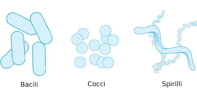
Even the shape of the bacterial colony grown on the nutrition medium also differs for different strains of bacteria.
Hence, under the microscope, the bacterial colony radius and shape can be viewed, and the strain of bacteria can be identified.
c. By extra-cellular characters
- Some extracellular characters are used to identify bacteria.
- Some bacteria have flagella for motion.
- So, they can be identified by the number and arrangement of flagella on the bacterial surface.
Ex: Atrichous bacteria have no flagella, Monotrichous have one flagellum, and Polytrichous have many flagella.
Even some bacteria have two flagella, one on each side of the cell.
2. By colony characteristics
- Here, bacterial cultures are identified as a whole.
- Since most bacteria grow in colonies and divide rapidly, they can be easily cultured on suitable nutrient media.
Based on the characteristics of the culture, they can be identified as
- The shape of culture: Circular, irregular, rhizoid, etc.
- Size of culture or colonies in millimeters.
- Type of elevation of culture, like effuse, convex, concave, etc.
- Margins of the colony, such as dentate, rough, smooth, etc.
- The surface of the colony: Smooth, wavy, papillate, etc.
- Color of culture.
3. Based on oxygen requirement
- Few bacteria are so specific in their oxygen or other requirements. Based on this specificity, they can be identified.
Ex: Aerobic bacteria require oxygen for survival.
- Anaerobic bacteria are killed when exposed to oxygen.
- Similarly, some require carbon dioxide, blood, or other pigments.
4. By differential staining
- The identification depends on the staining of bacteria.
Most bacteria can be stained with specific stains
Gram Staining:
- Gram-Positive: Thick peptidoglycan layer, retains crystal violet (purple).
- Gram-Negative: Thin peptidoglycan layer, outer membrane, counterstained pink/red by safranin.
Special Stains:
- Acid-Fast (Ziehl-Neelsen): Identifies Mycobacterium species (mycolic acid in cell wall).
- Capsule & Endospore Staining: Identifies protective capsules (S. pneumoniae) or dormant endospores (Bacillus, Clostridium).
5. Through biochemical tests.
This is one of the most widely used methods, which relies on bacterial metabolic characteristics.
a. Sugar fermentation test:
Bacteria are grown in a sugar medium, and the acid produced is indicated by a change in the medium’s color to red.
b. Litmus milk test:
As the name suggests, this test relies on changes in pH on litmus paper.
When bacteria are grown in this medium, there may be the production of acids or alkalis, or even no change in pH.
This change is dependent on the type of bacteria.
c. Indole production test
Bacteria are grown in the peptone water culture.
After 48 to 96 hours of incubation at 37°C, it is checked for the presence of red color.
The red color indicates the production of indole from the amino acid tryptophan.
The reagent used is called Kovac’s reagent.
d. Methyl Red test
Bacteria are grown in a glucose phosphate medium at 30°C for five days. Then, a few drops of 0.04% methyl red are added and mixed to observe for a color change.
The red color indicates a positive test for glucose-fermenting bacteria, while the yellow color indicates a negative test.
e. Citrate utilization test
The citrate medium, called Koser’s medium, is used here.
Citrate is the sole source of carbon, and its utilization is monitored by turbidity.
This test helps identify mostly intestinal gram-negative bacteria.
If positive, it indicates the above-said microbes. See in detail the citrate test.
f. Voges Proskauer Test
Here, 1 ml of glucose phosphate medium, 0.6 ml of a 5% α-naphthol solution in ethanol, and 0.2 ml of 40% KOH are added.
Acetyl methylcarbinol is produced from pyruvic acid, which is oxidized to diacetyl form to give a red color.
This test is used to detect bacteria such as Enterobacter and Klebsiella. See more details on the VP test.
- Catalase Test: Distinguishes Staphylococcus (+) from Streptococcus (-).
- Coagulase Test: Distinguishes Staphylococcus aureus (+) from Coagulase-Negative Staphylococci (CoNS) like S. epidermidis (-).
- Oxidase Test: Distinguishes Non-fermenters like Pseudomonas aeruginosa (+) from Enterobacteriaceae (-).
- IMViC Series (Indole, Methyl Red, VP, Citrate): Classic diagnostic panel for differentiating Gram-negative enteric bacilli (e.g., E. coli vs. Klebsiella).
6. Based on resistance
Antibiotics are specific to certain bacteria. Hence, a few bacteria develop resistance to certain types of antibiotics.
By adding antibiotics to the culture and measuring the microbe’s resistance to each antibiotic, the type of bacterium can be identified.
Ex: TB bacteria, i.e., Mycobacterium tuberculosis, are not affected by penicillin but killed by ciprofloxacin.
So, this differentiation can be used to identify a few bacteria.
7. Serological methods.
Here, bacterial identification is performed using antibodies and antigens specific to the suspected bacteria.
Antigens and antibodies are very specific and bind to a single type of bacteria.
8. By protein and nucleotide analysis.
The cellular constituents, such as protein content and DNA nucleotide sequence, are used to identify bacteria.
This requires methods such as polymerase chain reaction (PCR), gel electrophoresis, and radioimmunoassay.
9. Modern & Molecular Identification
PCR & Nucleic Acid Amplification (NAAT): Rapid detection of specific resistance genes or fastidious organisms.
MALDI-TOF MS (Mass Spectrometry): Rapid protein profiling using laser desorption; modern gold standard for clinical microbiology labs.
16S rRNA Gene Sequencing: Universal gene sequencing used for definitive taxonomic classification and identifying unculturable or rare bacteria.
Importance of bacterial Identification
1. Identify the disease
It helps to know the type of infection and the disease it causes.
For example, an acid-fast stain test for Mycobacterium can identify Tuberculosis.
So, bacterial identification is important in health care.
2. Select a suitable drug
Not all drugs (antibiotics) are active against all bacteria.
So, identification helps to target that specific bacteria with a suitable choice of drugs.
For example, rifamycin is best suited for tuberculosis and cannot be controlled by other antibiotics.
3. Evaluation of treatment progress
Identification of bacteria is also necessary to determine how effective the drug is and whether the patient is free of the said bacteria after treatment.
During treatment, the patient’s body samples are repeatedly checked to assess recovery.
This is ascertained by performing an identification test for the causative bacteria. A negative test result indicates he is free from infection.
4. For industrial purposes
It also helps isolate a specific strain of bacteria needed for research or other industrial processes, such as fermentation.
Many substances (vitamin B12), hormones (insulin), and supplements are produced using rDNA technology based on the principles of biotechnology.
5. For storage
Some bacteria are specifically stored for future use. Identification is also necessary to store the bacteria in pure form, free of foreign contamination.
These stored bacteria are supplied when required for research or industrial manufacture.
Because of the above advantages, bacterial identification is of utmost importance in microbiology.
Unlike viruses, bacteria are easily traced using simple staining methods.
Further, using a common microscope, one can even see whether they are moving (motile) or not, grouping, etc.
Identification of bacteria is a part of the diagnosis of many diseases and, hence, is in high demand for microbiologists.

I’ve been doing some reading about buffered peptone water, and I wanted to do a little more research. I’m glad you talked about how peptone water can grow bacteria. I’m going to have to look into some different buffered peptone water and see what I can find!