Microvilli were first seen by Granger and Baker in 1950 on intestinal epithelial cells by electron microscopy.
They appear as brush bristles on the apical cell surface of some epithelial cells in eukaryotes.
They may be present independently or in conjunction with villi.
Structure of microvilli
Microvilli are composed of microfilaments (actin) and cytoplasm and are covered by a plasma membrane. They do not have basal bodies.
Each microvillus has a structural core consisting of a bundle of cross-linked actin filaments.
About 20 to 30 densely bound actin filaments are cross-linked by bundling proteins espin, fimbrin and villin to form the core. They largely increase the cell’s surface area, thus increasing the absorption rate.
Microvilli have different dimensions on the basis of where they are found. But they are approximately 1 to 2 μm in length and about 90nm in diameter.
Microvilli are covered with glycocalyx, consisting of glycoproteins that are attached to a plasma membrane via transmembrane proteins.
Substances like the nutrients that need to be taken up by cells are bound to this layer. This layer also provides protection.
Gap junctions are present in the area of contact between the terminals of oocyte microvilli.
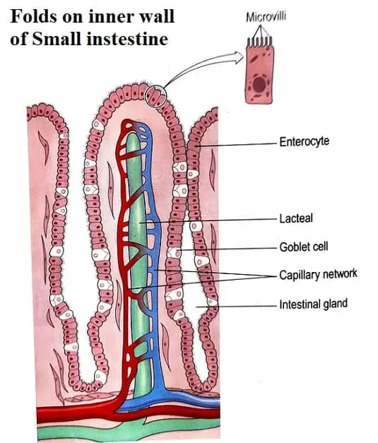
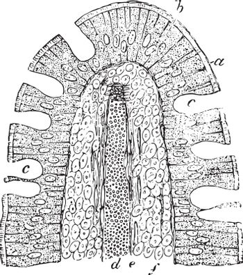
Microvilli structure increases the surface area to volume ratio. The small intestine wall is covered by several folds of mucus membrane called plicae circulares. The surface of these folds contains tiny projections called villi, which in turn have microvilli.
They increase the total area for absorption. The small intestine villi are covered by a single layer of simple columnar cells called goblet cells.
Goblet cells are scattered among the epithelial cells covering the villi and secrete mucin (which forms mucus). The goblet cells contain many microvilli.
Microvilli location
Microvilli are found in small intestinal epithelial cells and kidneys.
In the intestine, each villus is composed of many cells and each villus has thousands of microvilli on the apical surface forming a brush border.
They are also present on the plasma surface of the ovum to help in the anchoring of sperms.
Microvilli in the kidney (nephron)
Microvilli are present in the proximal convoluted tubule in the nephrons.
This part of the nephron has simple tall cuboidal epithelium.
The microvilli increase the surface area by 30-40 fold; thus, maximal water and sodium reabsorption occur here.
Microvilli in the digestive system.
The microvilli help digest and absorb intestinal contents by increasing the absorbing surface area.
They secrete brush border enzymes like disaccharidase and peptidase that hydrolyze disaccharides to monosaccharides and polypeptides to amino acids.
Brush border enzymes are the enzymes contained within the microvilli.
The microvillus membrane has transport proteins that help absorb sodium ions, glucose, and amino acids.
Receptors of certain substances are found on the microvilli at different levels in the small intestine.
This is why there is selective absorption of particular substances at particular sites like intrinsic-factor-bound vitamin B12 in the terminal ileum; iron and calcium in the duodenum and upper jejunum.
Actin and myosin are found in the microvilli, which have contractility functions.
Thus, the microvilli have a motor activity that is thought to initiate the small intestine’s mixing actions.
Microvilli in the respiratory tract
Microvilli are present on cells present between the ciliated columnar epithelial cells in the respiratory epithelium of the nasal cavity, larynx, trachea, and bronchi. They are also found in alveoli in diseased conditions.
Microvilli in neuron
Microvilli can arise at their apical side in some sensory neurons to form the sensory organelle.
This was especially observed in hair cells of the inner ear.
Microvilli in the uterus
The apical surface of the uterine epithelial cells is covered with microvilli which can vary in length and number with the menstrual cycle and during pregnancy.
They are under hormonal control. They are covered by glycocalyx and made of a mesh of actin filaments.
Microvilli in the tongue
Microvilli are located at the surface of taste buds in the taste pores.
They detect dissolved chemicals in food, which activate receptor cells in the taste buds. The epithelial cells of taste buds are called gustatory cells.
Microvilli in neutrophil
The microvilli help the neutrophil roll on the endothelium in case of inflammatory reactions.
Microvilli in the liver
The epithelial cells of the liver (hepatocytes) have many microvilli on their surface, which project into perisinusoidal space in the liver and help in absorption from plasma.
Microvilli Function and Significance
Microvilli largely increase the surface area of the cell. It increases the surface of nutrient absorption in the gastrointestinal tract and kidney.
The microvillar membrane is also packed with enzymes that work in the breakdown of complex nutrients into simpler compounds that can be absorbed, e.g., there are high amounts of glycosidases on small intestinal microvilli.
Thus, microvilli increase the absorption of substances by both increasing surface area and digestive enzymes.
In some cases, they also help in secretion, cellular adhesion, and mechano-transduction.
Microvilli resist the binding of microbes like bacteria via electrostatic repulsions.
Microvilli increase active transport by increasing the surface area so more transporters can be present per cell.
Thus more transporters use more ATP to transport more electrolytes or nutrients.
| Characters | Microvilli | Cilia |
|---|---|---|
| Appearance | These are microscopic cellular membrane projections that are non-motile | These are fine hair-like cellular membrane projections that move back and forth. |
| Structure | microvilli are shorter and thicker and covered by glycocalyx | cilia that arise from basal granules and show (9+2) arrangement in ultrastructure. |
| Shape | Microvilli are cylindrical and have blunt ends. | Cilia are cylindrical with tapering ends. |
| Location | Microvilli are found mostly in the columnar epithelium of the small intestine and kidney | cilia are found in the respiratory and uterine tract. |
| Function | They increase the surface area and help in adsorption | They help move fluid in one specific direction |
Microvilli Repair and Regeneration
Microvilli in the gut and other lining get damaged due to sloughing of cells, actions of toxins, stresses, etc. Repair of microvilli occurs via intrinsic reparative processes.
The entire cell is not regenerated and replaced every time.
Microvilli Disease
A disease of the microvilli affects infants and children called Microvilli Inclusion Disease (MVID).
The patient cannot digest the food properly and cannot absorb nutrients effectively. It is characterized by chronic, watery diarrhea.
Are microvilli cells?
Microvilli are not cells. They are parts of cells. They are outgrowths from the cell surface.
Are microvilli found in plant cells?
Microvilli are present in the root hair cells of plant cells. This increases the surface area for increased diffusion and osmosis to absorb more water and nutrients from the soil.
Are microvilli found in prokaryotes?
Microvilli are not present in prokaryotes.
Are microvilli anchored by desmosomes?
No, but the desmosome protein desmoplakin affects the length of microvilli.
Can microvilli regenerate?
Yes, microvilli can regenerate by the repair process
Do microvilli contain cytoskeleton elements?
Yes, microvilli consist of cytoskeleton elements made up of a network of actin filaments.
Does the stomach have microvilli?
No, the stomach does not have microvilli.
What is the functional difference between microvilli and cilia
Microvilli are meant for absorption of substances while cilia are meant to move the substances in a particular direction.
