The kidney is the primary organ of excretion in the human body.
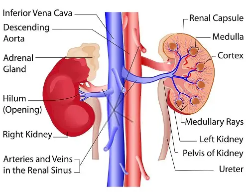
A pair of kidneys is attached to the posterior abdominal wall. Each lies on either side of the vertebral column.
Gross Structure Of the Kidney
- A kidney is a bean-shaped organ and is reddish-brown in color.
- It has a dimension of 11 cm in length, 6 cm in width, and 3 cm in thickness.
- The average kidney weighs about 150 grams in males and 135 grams in females.
Kidney Structure by longitudinal section
The kidney has 3 several layers of different tissues, which have different functions. These include
- Outer fibrous capsule
- The Cortex
- Medulla in the center.
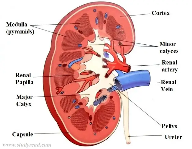
Fibrous capsule (renal capsule)
- This is the outermost cover of the kidney.
- It surrounds the kidney and helps to enclose the internal tissues.
Cortex
- This is the peripheral region of the kidney lying immediately below the kidney.
- It is also called the renal cortex.
- Cortex is highly vascularized and hence is reddish-brown in color.
The renal cortex has two parts:
- The Cortical arches or lobules, which form arches over the pyramids.
- The renal columns, which run between the pyramids.
Medulla
- This is the innermost layer of the kidney.
- It is also called the Renal Medulla.
- It has pale-colored conical striations called pyramids.
- Each kidney has 9 to 10 renal pyramids.
The medulla is comparatively less vascularized than the cortex and hence appears pale to the naked eye.
The lobe of the kidney is formed by each pyramid and its overlying cortex.
Sinus
- Within the renal medulla is an open space, called the sinus, or renal sinus.
- These sinuses are the spaces that run from the hilum of the kidney up to the kidney’s functional parts.
Renal sinuses contain
- Renal Artery and its branches
- The tributaries of the renal vein
- Minor and Major and calyces.
These major calyces are the branchings of the renal pelvis.
- The renal pelvis divides into 2 to 3 major renal calyces. Each major calyces divide into 7 to 13 minor calyces.
- The urine from the pyramid reaches minor calyces and then into major calyces. From there, it passes into the renal pelvis.
- The renal pelvis opens into the ureters.
- The walls of the renal pelvis are made of smooth muscle with a lining of epithelium.
- The peristaltic movements originating in the smooth muscle cells of the pelvis proper propel the urine into the ureters.
Blood Supply of the Kidney
Kidneys receive blood from the Renal Arteries, and are direct branches of the Abdominal Aorta.
The contents of the renal hilum, the entry point of the Kidney, which are devoid of the renal capsule, are
- Renal Artery
- Renal vein
- Nerves
- Lymphatics
- Ureter
Nephron and its structure
A nephron is the functional and structural unit of the kidneys.
- Each kidney has 1 to 2 million functional nephrons.
- But the number of collecting ducts is fewer.
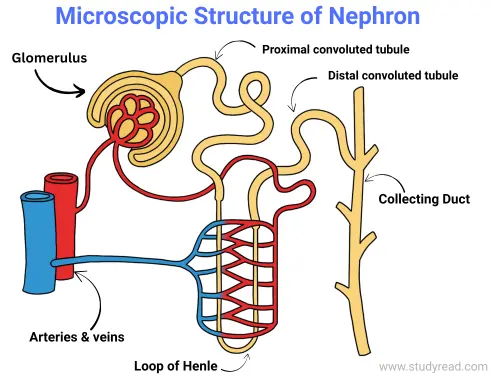
Each Nephron has four parts
I. Bowman’s Capsule
2. Proximal Convoluted Tubule (PCT)
3. Loop of Henle; That again has two parts
- Ascending loop of Henle
- Descending loop of Henle
4. Distal Convoluted Tubule (DCT).
👉 Also Collecting Ducts (CT):
- These are tubules where the distal convoluted tubules empty their contents.
- These CTs act as a common tube for many nephrons.
Bowman’s capsule
- Bowman’s capsule is the thin-layered, unilamellar-lined, closed chamber enclosing the glomerulus and continuing with the proximal convoluted Tubule.
- It encloses a glomerulus, which is a tuft of capillaries arising from the afferent arteriole and emerging out of the glomerulus as the efferent arteriole.
Capillaries are one cell layer in thickness, and the diameter of the afferent arteriole is twice the diameter of the efferent arteriole.
This creates pressure rises inside the glomerulus, which pushes the plasma to filter along with its contents, like
- The glucose,
- Ions,
- Excretory products like urea and creatinine.
This filtrate is called the primary filtrate.
An afferent arteriole is a branch of an interlobular artery, which is a branch of a renal artery that branches into the glomerular capillaries.
These capillaries reunite to form the efferent arteriole, which then again divides into peritubular capillaries.
Peritubular Capillaries are found only in the case of cortical Nephrons.
In the case of Juxtaglomerular nephrons, they are straight and parallel to the loop of Henle, called vasa recta.
Special types of cells are found in Bowman’s capsule called podocytes, which have gaps in between them to facilitate this ultrafiltration.
Proximal Convoluted Tubule
- Proximal Convoluted Tubule (PCT) has an average adult length of 15mm, with a 55micron diameter.
- It is highly coiled and surrounds the glomerular capsule.
- Proximal Convoluted Tubule reabsorbs about 67% of the filtrate, i.e., two-thirds of the primary filtrate enters the blood.
The cells of the PCT have ion channels, like the
- Sodium-potassium cotransporter,
- Divalent ion transporter,
- Glucose absorbers
which actively reabsorb ions and glucose (100%) from the primary filtrate.
- Passive reabsorption of water happens due to osmotic pressure differences.
Loop of Henle
The loop of Henle is the site of absorption for
- urea
- Ions and
- water
According to the body’s needs.
Distal Convoluted Tubule
- The Distal Convoluted Tubule (DCT) is a continuation of the Loop of Henle.
- The surface cells help in the absorption of potassium ions and water according to the body’s needs.
Collecting Duct
- Collecting Ducts are the successors of the Distal Convoluted Tubule.
- It has type I and P cells that actively secrete bicarbonate ions and urea into the fluid.
- Besides, there is further water reabsorption according to need.
- The fluid leaving the collecting ducts is the final composition of urine.
- It is highly concentrated than the primary filtrate and is completely sterile.
The urine is collected by the ureter and carried forward towards the urinary bladder.
It is then excreted outside the body via the urethra.
Nephrons are of two types:
1. Cortical Nephron
- These Nephrons constitute about 85% of all the Nephrons in the kidney.
- They are smaller in size with a shorter loop of Henle and penetrate less into the medulla.
- It is more confined in the cortical region of the kidney. Their glomerulus is located in the superficial parts of the renal cortex.
2. Juxtaglomerular Nephrons
- These are the Nephrons, which constitute the remaining 15% of the Nephrons present in the kidney.
- They are larger in size, with a longer loop of Henle going deep down up to the renal Medulla.
- They have their glomerulus located in the deeper part of the renal cortex.
Functions of the kidneys
- The kidneys maintain the concentration of blood in our body.
- They also help regulate blood pH and electrolyte levels.
- They form the major organs of excretion in the whole excretory process.
- Kidneys secrete hormones renin and erythropoietin.
- Renin helps in the maintenance of blood pressure and volume.
- Erythropoietin boosts the production of RBCs under hypoxia.
